EP2348052A2 - Internalizing human monoclonal antibodies targeting prostate cancer cells in situ - Google Patents
Internalizing human monoclonal antibodies targeting prostate cancer cells in situ Download PDFInfo
- Publication number
- EP2348052A2 EP2348052A2 EP11159671A EP11159671A EP2348052A2 EP 2348052 A2 EP2348052 A2 EP 2348052A2 EP 11159671 A EP11159671 A EP 11159671A EP 11159671 A EP11159671 A EP 11159671A EP 2348052 A2 EP2348052 A2 EP 2348052A2
- Authority
- EP
- European Patent Office
- Prior art keywords
- antibody
- seq
- antibodies
- scfv
- cells
- Prior art date
- Legal status (The legal status is an assumption and is not a legal conclusion. Google has not performed a legal analysis and makes no representation as to the accuracy of the status listed.)
- Withdrawn
Links
Images


Classifications
-
- C—CHEMISTRY; METALLURGY
- C07—ORGANIC CHEMISTRY
- C07K—PEPTIDES
- C07K16/00—Immunoglobulins [IGs], e.g. monoclonal or polyclonal antibodies
- C07K16/18—Immunoglobulins [IGs], e.g. monoclonal or polyclonal antibodies against material from animals or humans
- C07K16/28—Immunoglobulins [IGs], e.g. monoclonal or polyclonal antibodies against material from animals or humans against receptors, cell surface antigens or cell surface determinants
- C07K16/30—Immunoglobulins [IGs], e.g. monoclonal or polyclonal antibodies against material from animals or humans against receptors, cell surface antigens or cell surface determinants from tumour cells
- C07K16/3069—Reproductive system, e.g. ovaria, uterus, testes, prostate
-
- A—HUMAN NECESSITIES
- A61—MEDICAL OR VETERINARY SCIENCE; HYGIENE
- A61K—PREPARATIONS FOR MEDICAL, DENTAL OR TOILETRY PURPOSES
- A61K51/00—Preparations containing radioactive substances for use in therapy or testing in vivo
- A61K51/02—Preparations containing radioactive substances for use in therapy or testing in vivo characterised by the carrier, i.e. characterised by the agent or material covalently linked or complexing the radioactive nucleus
- A61K51/04—Organic compounds
- A61K51/08—Peptides, e.g. proteins, carriers being peptides, polyamino acids, proteins
- A61K51/10—Antibodies or immunoglobulins; Fragments thereof, the carrier being an antibody, an immunoglobulin or a fragment thereof, e.g. a camelised human single domain antibody or the Fc fragment of an antibody
- A61K51/1045—Antibodies or immunoglobulins; Fragments thereof, the carrier being an antibody, an immunoglobulin or a fragment thereof, e.g. a camelised human single domain antibody or the Fc fragment of an antibody against animal or human tumor cells or tumor cell determinants
- A61K51/1072—Antibodies or immunoglobulins; Fragments thereof, the carrier being an antibody, an immunoglobulin or a fragment thereof, e.g. a camelised human single domain antibody or the Fc fragment of an antibody against animal or human tumor cells or tumor cell determinants the tumor cell being from the reproductive system, e.g. ovaria, uterus, testes or prostate
-
- A—HUMAN NECESSITIES
- A61—MEDICAL OR VETERINARY SCIENCE; HYGIENE
- A61P—SPECIFIC THERAPEUTIC ACTIVITY OF CHEMICAL COMPOUNDS OR MEDICINAL PREPARATIONS
- A61P35/00—Antineoplastic agents
-
- C—CHEMISTRY; METALLURGY
- C07—ORGANIC CHEMISTRY
- C07K—PEPTIDES
- C07K16/00—Immunoglobulins [IGs], e.g. monoclonal or polyclonal antibodies
- C07K16/005—Immunoglobulins [IGs], e.g. monoclonal or polyclonal antibodies constructed by phage libraries
-
- C—CHEMISTRY; METALLURGY
- C07—ORGANIC CHEMISTRY
- C07K—PEPTIDES
- C07K16/00—Immunoglobulins [IGs], e.g. monoclonal or polyclonal antibodies
- C07K16/18—Immunoglobulins [IGs], e.g. monoclonal or polyclonal antibodies against material from animals or humans
-
- C—CHEMISTRY; METALLURGY
- C07—ORGANIC CHEMISTRY
- C07K—PEPTIDES
- C07K16/00—Immunoglobulins [IGs], e.g. monoclonal or polyclonal antibodies
- C07K16/18—Immunoglobulins [IGs], e.g. monoclonal or polyclonal antibodies against material from animals or humans
- C07K16/28—Immunoglobulins [IGs], e.g. monoclonal or polyclonal antibodies against material from animals or humans against receptors, cell surface antigens or cell surface determinants
- C07K16/2803—Immunoglobulins [IGs], e.g. monoclonal or polyclonal antibodies against material from animals or humans against receptors, cell surface antigens or cell surface determinants against the immunoglobulin superfamily
-
- C—CHEMISTRY; METALLURGY
- C07—ORGANIC CHEMISTRY
- C07K—PEPTIDES
- C07K16/00—Immunoglobulins [IGs], e.g. monoclonal or polyclonal antibodies
- C07K16/18—Immunoglobulins [IGs], e.g. monoclonal or polyclonal antibodies against material from animals or humans
- C07K16/28—Immunoglobulins [IGs], e.g. monoclonal or polyclonal antibodies against material from animals or humans against receptors, cell surface antigens or cell surface determinants
- C07K16/30—Immunoglobulins [IGs], e.g. monoclonal or polyclonal antibodies against material from animals or humans against receptors, cell surface antigens or cell surface determinants from tumour cells
-
- G—PHYSICS
- G01—MEASURING; TESTING
- G01N—INVESTIGATING OR ANALYSING MATERIALS BY DETERMINING THEIR CHEMICAL OR PHYSICAL PROPERTIES
- G01N33/00—Investigating or analysing materials by specific methods not covered by groups G01N1/00 - G01N31/00
- G01N33/48—Biological material, e.g. blood, urine; Haemocytometers
- G01N33/50—Chemical analysis of biological material, e.g. blood, urine; Testing involving biospecific ligand binding methods; Immunological testing
- G01N33/5005—Chemical analysis of biological material, e.g. blood, urine; Testing involving biospecific ligand binding methods; Immunological testing involving human or animal cells
- G01N33/5008—Chemical analysis of biological material, e.g. blood, urine; Testing involving biospecific ligand binding methods; Immunological testing involving human or animal cells for testing or evaluating the effect of chemical or biological compounds, e.g. drugs, cosmetics
- G01N33/5044—Chemical analysis of biological material, e.g. blood, urine; Testing involving biospecific ligand binding methods; Immunological testing involving human or animal cells for testing or evaluating the effect of chemical or biological compounds, e.g. drugs, cosmetics involving specific cell types
-
- G—PHYSICS
- G01—MEASURING; TESTING
- G01N—INVESTIGATING OR ANALYSING MATERIALS BY DETERMINING THEIR CHEMICAL OR PHYSICAL PROPERTIES
- G01N33/00—Investigating or analysing materials by specific methods not covered by groups G01N1/00 - G01N31/00
- G01N33/48—Biological material, e.g. blood, urine; Haemocytometers
- G01N33/50—Chemical analysis of biological material, e.g. blood, urine; Testing involving biospecific ligand binding methods; Immunological testing
- G01N33/53—Immunoassay; Biospecific binding assay; Materials therefor
- G01N33/574—Immunoassay; Biospecific binding assay; Materials therefor for cancer
- G01N33/57407—Specifically defined cancers
- G01N33/57434—Specifically defined cancers of prostate
-
- G—PHYSICS
- G01—MEASURING; TESTING
- G01N—INVESTIGATING OR ANALYSING MATERIALS BY DETERMINING THEIR CHEMICAL OR PHYSICAL PROPERTIES
- G01N33/00—Investigating or analysing materials by specific methods not covered by groups G01N1/00 - G01N31/00
- G01N33/48—Biological material, e.g. blood, urine; Haemocytometers
- G01N33/50—Chemical analysis of biological material, e.g. blood, urine; Testing involving biospecific ligand binding methods; Immunological testing
- G01N33/68—Chemical analysis of biological material, e.g. blood, urine; Testing involving biospecific ligand binding methods; Immunological testing involving proteins, peptides or amino acids
- G01N33/6803—General methods of protein analysis not limited to specific proteins or families of proteins
- G01N33/6845—Methods of identifying protein-protein interactions in protein mixtures
-
- G—PHYSICS
- G01—MEASURING; TESTING
- G01N—INVESTIGATING OR ANALYSING MATERIALS BY DETERMINING THEIR CHEMICAL OR PHYSICAL PROPERTIES
- G01N33/00—Investigating or analysing materials by specific methods not covered by groups G01N1/00 - G01N31/00
- G01N33/48—Biological material, e.g. blood, urine; Haemocytometers
- G01N33/50—Chemical analysis of biological material, e.g. blood, urine; Testing involving biospecific ligand binding methods; Immunological testing
- G01N33/68—Chemical analysis of biological material, e.g. blood, urine; Testing involving biospecific ligand binding methods; Immunological testing involving proteins, peptides or amino acids
- G01N33/6854—Immunoglobulins
-
- A—HUMAN NECESSITIES
- A61—MEDICAL OR VETERINARY SCIENCE; HYGIENE
- A61K—PREPARATIONS FOR MEDICAL, DENTAL OR TOILETRY PURPOSES
- A61K39/00—Medicinal preparations containing antigens or antibodies
- A61K2039/505—Medicinal preparations containing antigens or antibodies comprising antibodies
-
- C—CHEMISTRY; METALLURGY
- C07—ORGANIC CHEMISTRY
- C07K—PEPTIDES
- C07K2317/00—Immunoglobulins specific features
- C07K2317/20—Immunoglobulins specific features characterized by taxonomic origin
- C07K2317/21—Immunoglobulins specific features characterized by taxonomic origin from primates, e.g. man
-
- C—CHEMISTRY; METALLURGY
- C07—ORGANIC CHEMISTRY
- C07K—PEPTIDES
- C07K2317/00—Immunoglobulins specific features
- C07K2317/50—Immunoglobulins specific features characterized by immunoglobulin fragments
- C07K2317/56—Immunoglobulins specific features characterized by immunoglobulin fragments variable (Fv) region, i.e. VH and/or VL
- C07K2317/565—Complementarity determining region [CDR]
-
- C—CHEMISTRY; METALLURGY
- C07—ORGANIC CHEMISTRY
- C07K—PEPTIDES
- C07K2317/00—Immunoglobulins specific features
- C07K2317/50—Immunoglobulins specific features characterized by immunoglobulin fragments
- C07K2317/56—Immunoglobulins specific features characterized by immunoglobulin fragments variable (Fv) region, i.e. VH and/or VL
- C07K2317/569—Single domain, e.g. dAb, sdAb, VHH, VNAR or nanobody®
-
- C—CHEMISTRY; METALLURGY
- C07—ORGANIC CHEMISTRY
- C07K—PEPTIDES
- C07K2317/00—Immunoglobulins specific features
- C07K2317/60—Immunoglobulins specific features characterized by non-natural combinations of immunoglobulin fragments
- C07K2317/62—Immunoglobulins specific features characterized by non-natural combinations of immunoglobulin fragments comprising only variable region components
- C07K2317/622—Single chain antibody (scFv)
-
- C—CHEMISTRY; METALLURGY
- C07—ORGANIC CHEMISTRY
- C07K—PEPTIDES
- C07K2317/00—Immunoglobulins specific features
- C07K2317/70—Immunoglobulins specific features characterized by effect upon binding to a cell or to an antigen
- C07K2317/77—Internalization into the cell
Definitions
- This invention pertains to the field of antibodies, immunodiagnostics, and immunotherapeutics.
- this invention pertains to novel methods for in vivo screening of antibodies and the discovery of prostate cancer specific internalizing antibodies using such methods.
- tumor cell surface antigens are invaluable targets for therapeutic development.
- the epitope space at the cell surface is highly complex.
- Relevant antigens may include glycosylated proteins and other post-translationally modified products that may not be readily predicted from studies of genomic copy number or mRNA expression levels ( Liu et al. (2004) Cancer Res. 64: 704-710 ; Kobata and Amano (2005) Immunol. Cell Biol. 83: 429-439 ; Birkle et al. (2003) Biochimie (Paris) 85: 455-463 ; Hakomori (2001) Adv. Exp. Med. Biol. 491: 369-402 ; Hanisch, F. G. (2001) O-Glycosylation of the mucin type. Biol. Chem. 382, 143-1 49 ; Ugorski and Laskowska (2002) Acta Biochim. Pol. 49:303-311 ).
- mAbs monoclonal antibodies
- mAbs monoclonal antibodies
- they can be used to efficiently map the tumor cell surface epitope space ( Liu et al. (2004) Cancer Res. 64, 704-710 ). Isolating these epitopes enables the antibodies to achieve specific binding to neoplastic cells, an ability that could be utilized in applications such as induction of antibody-dependent cell cytotoxicity ( Clynes et al. (2000) Nat. Med. 6: 443-446 ) or inhibition of signaling pathways involved in tumor cell migration, growth, and survival ( McWhirter et al. (2006) Proc. Natl. Acad.
- Phage antibody display has been widely used to develop cancer-specific antibodies ( Liu et al. (2004) Cancer Res. 64: 704-710 ; Liu and Marks (2000) Anal. Biochem. 286: 119-128 ; 15. Marks et al. (1992) Biotechnology (N. Y.) 10: 779-783 ; Marks et al. (1991) J. Mol. Biol. 222: 581-597 ; Marks et al. (1992) J. Biol. Chem. 267: 16007-16010 ; Sharon et al. (2005) J. Cell. Biochem. 96: 305-313 ; Silacci et al. (2005) Proteomics 5: 2340 -2350 ; Gao et al.
- a combinatorial phage antibody library serves as a source of random shape repertoire that can be used to probe neoplastic variations on the surface of cancer cells ( Liu et al. (2004) Cancer Res. 64: 704-710 ; Geuijen et al. (2005) Eur. J.
- cancer cell lines may express cell surface epitopes that differ from those present in the original cancerous tissue. Tissue sections from cancer patients would be an ideal selection target in the development of cancer-specific antibodies; however, most tissues taken during surgeries, biopsies, or autopsies are composed of heterogeneous cell populations. This seemingly poses a serious obstacle to selection methods that would specifically target cancer cells in tissue.
- this invention pertains to the development of a method that allows selection of antibodies against tumor cells in situ using laser capture microdissection. By restricting antibody selection to binders of internalizing epitopes, a panel of phage antibodies that target clinically represented prostate cancer antigens was generated.
- this invention provides an isolated antibody that specifically binds and, optionally, is internalized into a prostate cancer cell.
- the antibody is an antibody that specifically binds to an epitope that is specifically bound by bound an antibody selected from the group consisting of e.g., 3051.1, G12FC3, M6c42b, 4F3YW, M40pr146, UA20, UA8, 585II41, 585II41.1, 585II56, 3076, 3051, M49R, RCI-14, II79_4, II79_3, T5II-4B.1, T5II-4B.2, RCI-11, RCI-20, CI-11A, CI-14A, and/or S95-2.
- the antibody comprises one, two, or three complementarity determining regions (CDRs of the variable light (VL domain of an antibody selected from the group consisting of e.g., 3051.1, G12FC3, M6c42b, 4F3YW, M40pr146, UA20, UA8, 585II41, 585II41.1, 585II56, 3076, 3051, M49R, RCI-14, II79_4, II79_3, T5II-4B.1, T5II-4B.2, RCI-11, RCI-20, CI-11A, CI-14A, and/or S95-2; and/or one, two, or three complementarity determining regions (CDRs of the variable heavy (VH domain of an antibody selected from the group consisting of e.g.
- the antibody comprises the three VH CDRs and/or the three VL CDRs of an antibody selected from the group consisting of e.g., 3051.1, G12FC3, M6c42b, 4F3YW, M40pr146, UA20, UA8, 585II41, 585II41.1, 585II56, 3076, 3051, M49R, RCI-14, II79_4, II79_3, T5II-4B.1, T511-4B.2, RCI-11, RCI-20, CI-11A, CI-14A, and/or S95-2.
- an antibody selected from the group consisting of e.g., 3051.1, G12FC3, M6c42b, 4F3YW, M40pr146, UA20, UA8, 585II41, 585II41.1, 585II56, 3076, 3051, M49R, RCI-14, II79_4, II79_3, T5II-4B.1, T511-4B.2,
- the antibody comprises the VH domain and/or the VL domain of an antibody selected from the group consisting of e.g., 3051.1, G12FC3, M6c42b, 4F3YW, M40pr146, UA20, UA8, 585II41, 585II41.1, 585II56, 3076, 3051, M49R, RCI-14, II79_4, II79_3, T511-4B.1, T511-4B.2, RCI-11, RCI-20, CI-11A, CI-14A, and/or S95-2.
- the antibody is a single chain antibody.
- the VL region is attached to the VH region by a (Gly 4 Ser 3 SEQ ID NO:(SEQ ID NO:1) linker.
- the antibody is an intact full antibody, a Fab, an (Fab') 2 , an scFv, and an (ScFv'0 2 , a unibody, or an affibody.
- the antibody comprises a diabody.
- the antibody is a single chain antibody selected from the group consisting of 3051.1, G12FC3, M6c42b, 4F3YW, M40pr146, UA20, UAB, 585II41, 585II41.1, 585II56, 3076, 3051, M49R, RCI-14, II79_4, II79_3, T5II-4B.1, T5II-4B.2, RCI-11, RCI-20, CI-11A, CI-14A, and S95-2.
- chimeric moieties comprising an effector attached to any one or more of the antibodies described herein.
- the effector is selected from the group consisting of an epitope tag, a second antibody, a label, a cytotoxin, a liposome, a radionuclide, a drug, a prodrug, a viral particle, a cytokine, and a chelate.
- the effector comprises an epitope tag selected from the group consisting of an avidin, and a biotin.
- the effector comprises a cytotoxin selected from the group consisting of a Diphtheria toxin, a Pseudomonas exotoxin, a ricin, an abrin, and a thymidine kinase.
- the effector comprises a chelate comprising a metal isotope selected from the group consisting of 99 Tc, 203 Pb, 67 Ga, 68 Ga, 72 As, 111 In, 113 In, 97 Ru, 62 Cu, 641 Cu, 52 Fe, 52 Mn, 51 Cr, 186 , Re, 188 Re, 77 As, 90 Y, 67 Cu, 169 Er, 121 Sn, 127 Te, 142 Pr, 143 Pr, 198 Au, 199 Au, 161 Tb, 109 Pd, 165 Dy, 149 Pm 151 Pm, 153 Sm, 157 Gd, 159 Gd, 166 Ho, 172 Tm, 169 Yb 175 Yb, 177 LU, 105 Rh, and 111 Ag.
- the effector comprises an alpha emitter (e.g., bismuth 213).
- the effector is a chelate comprising DOTA.
- the effector comprises a metal isotope selected from the group
- compositions comprising a pharmaceutically acceptable excipient and an antibody or a chimeric moiety as described herein.
- the pharmaceutical formulation of claim is a unit dosage formulation.
- the pharmaceutical formulation is formulated for administration by a route such as intraperitoneal administration, intravenous injection, intramuscular injection, subcutaneous administration, direct administration to a tumor and/or surgical site, transcutaneous administration, subcutaneous depot formulation, oral administration, inhalation administration, rectal administration and the like.
- methods for delivering an effector to a prostate cancer cell.
- the methods typically involve administering to a cell, tissue, or organism, a composition comprising an antibody as described herein attached to an effector; whereby the antibody preferentially interacts with a prostate cancer cell thereby delivering the effector to the prostate cancer cell.
- the preferentially interacting comprises being internalized by said cancer cell.
- the effector comprises an anti-cancer agent and/or a detectable label.
- the administering comprises administering to a human or to a non-human mammal.
- the administering comprises administering parenterally.
- the administering comprises administering into a tumor or a surgical site.
- methods for inhibiting the growth or proliferation of a prostate cancer cell.
- the methods typically involve contacting the cancer cell with an antibody as described herein and/or with a chimeric moiety comprising one or more antibodies as described herein attached to an anti-cancer drug (e.g., a lipid complexed with an anti-cancer drug, a liposome containing an anti-cancer drug, etc .), or a radionuclide.
- an anti-cancer drug e.g., a lipid complexed with an anti-cancer drug, a liposome containing an anti-cancer drug, etc .
- a radionuclide e.g., a radionuclide.
- the cancer cell is a metastatic cell.
- the cancer cell is a solid tumor cell.
- methods are also provided for detecting a prostate cancer cell.
- the methods typically involve contacting the prostate cancer cell with a chimeric molecule comprising an antibody as described herein attached to a detectable label; and detecting the presence and/or location of said detectable label where the presence and/or location is an indicator of the location and/or presence of a prostate cancer cell.
- the detectable label is selected from the group consisting of a gamma-emitter, a positron-emitter, an x-ray emitter, an alpha emitter, and a fluorescence-emitter.
- the methods typically involve contacting a prostate cancer cell with a chimeric molecule comprising an antibody as described herein attached to an epitope tag; contacting the chimeric molecule with a chelate comprising a detectable moiety whereby the chelate binds to the epitope tag thereby associating said detectable moiety with the chelate; and detecting the detectable moiety where the presence and/or location of the detectable moiety is an indicator of the location and/or presence of a prostate cancer cell.
- the detectable moiety or detectable label is a radionuclide.
- the detectable moiety or detectable label is selected from the group consisting of a gamma-emitter, a positron-emitter, an alpha emitter, an x-ray emitter, and a fluorescence-emitter.
- the detecting comprises external imaging. In certain embodiments the detecting comprises internal imaging.
- the detectable moiety or detectable label comprises a metal isotope selected from the group consisting of to 99 Tc , 203 Pb, 67 Ga, 68 Ga, 72 As, 111 In, 113m In, 97 Ru, 62 Cu, 641 Cu , 52 Fe, 52m Mn, 51 Cr, 186 , Re, 188 Re, 77 As, 90 Y, 67 Cu, 169 Er, 121 Sn, 127 Te, 142 Pr, 143 Pr, 198 Au, 199 Au, 161 Tb, 109 Pd, 165 Dy, 149 Pm, 151 Pm, 153 Sm, 157 Gd, 159 Gd, 166 Ho, 172 Tm, 169 Yb, 175 Yb, 177 Lu, 99m Tc , 105 Rh, and 111 Ag.
- the chelate comprises DOTA.
- the epitope tag is an avidin or a biotin.
- nucleic acids encoding an antibody as described herein.
- the nucleic acids comprise a vector and can be present in a cell whereby the cell expresses the antibody.
- this invention provides methods of identifying an antibody that preferentially binds to and/or is internalized by a target cell type that expresses a marker in vivo.
- the methods typically involve providing a display library (e.g., a yeast- or phage-display library); contacting a tissue compromising the cell type with members of the library; isolating groups of cells from the tissue using laser capture microdissection; and recovering members of the library that bind to cells in the isolated groups.
- the recovering comprises identifying members of the library that are internalized into cells in the isolated groups.
- the target cell type is a pathological cell or a healthy cell characteristic of a particular tissue.
- the target cell type is a cancer cell (e.g., a cell of a cancer selected from the group consisting of a lung cancer, a bronchus cancer, a colorectal cancer, a prostate cancer, a breast cancer, a pancreas cancer, a stomach cancer, an ovarian cancer, a urinary bladder cancer, a brain or central nervous system cancer, a peripheral nervous system cancer, an esophageal cancer, a cervical cancer, a melanoma, a uterine or endometrial cancer, a cancer of the oral cavity or pharynx, a liver cancer, a kidney cancer, a biliary tract cancer, a small bowel or appendix cancer, a salivary gland cancer, a thyroid gland cancer, an adrenal gland cancer, an osteosarcoma, a chondrosarcoma, a liposarcoma, a testicular cancer, and a malignant fibrous histiocytoma).
- a cancer cell e
- the method involves counterselecting the library on a normal cell population to reduce or eliminate members of the library that bind to normal cells.
- the providing comprises preselecting the library on a panel of tumor cell lines to create a library enriched for binders to functional cell surface epitopes on tumor cells.
- the preselecting is under internalizing conditions.
- the recovering comprises amplifying a nucleic acid sequence encoding all or part of a displayed VH and/or VL domain from the bound or internalized members of the library.
- polypeptide polypeptide
- peptide protein
- protein protein
- amino acid polymers in which one or more amino acid residue is an artificial chemical analogue of a corresponding naturally occurring amino acid, as well as to naturally occurring amino acid polymers.
- amino acid polymers in which one or more amino acid residue is an artificial chemical analogue of a corresponding naturally occurring amino acid, as well as to naturally occurring amino acid polymers.
- the term also includes variants on the traditional peptide linkage joining the amino acids making up the polypeptide.
- nucleic acid or “oligonucleotide” or grammatical equivalents herein refer to at least two nucleotides covalently linked together.
- a nucleic acid of the present invention is preferably single-stranded or double stranded and will generally contain phosphodiester bonds, although in some cases, as outlined below, nucleic acid analogs are included that may have alternate backbones, comprising, for example, phosphoramide ( Beaucage et al. (1993) Tetrahedron 49(10):1925 ) and references therein; Letsinger (1970) J. Org. Chem. 35:3800 ; Sblul et al. (1977) Eur. J. Biochem.
- biotin refers to biotin and modified biotins or biotin analogues that are capable of binding avidin or various avidin analogues.
- Biotin can be, inter alia, modified by the addition of one or more addends, usually through its free carboxyl residue.
- Useful biotin derivatives include, but are not limited to, active esters, amines, hydrazides and thiol groups that are coupled with a complimentary reactive group such as an amine, an acyl or alkyl group, a carbonyl group, an alkyl halide or a Michael-type acceptor on the appended compound or polymer.
- Avidin typically found in egg whites, has a very high binding affinity for biotin, which is a B-complex vitamin ( Wilcheck et al. (1988) Anal. Biochem, 171: 1 ).
- Streptavidin derived from Streptomyces avidinii, is similar to avidin, but has lower nonspecific tissue binding, and therefore often is used in place of avidin.
- "avidin” includes all of its biological forms either in their natural states or in their modified forms. Modified forms of avidin which have been treated to remove the protein's carbohydrate residues (“deglycosylated avidin"), and/or its highly basic charge (“neutral avidin”), for example, also are useful in the invention. Both avidin and streptavidin have a tetravalency for biotin, thus permitting amplification when the former bind to biotin.
- four detection or therapeutic agents such as nuclides, can be attached to each targeting protein.
- residue refers to natural, synthetic, or modified amino acids.
- an “antibody” refers to a protein consisting of one or more polypeptides substantially encoded by immunoglobulin genes or fragments of immunoglobulin genes.
- the recognized immunoglobulin genes include the kappa, lambda, alpha, gamma, delta, epsilon and mu constant region genes, as well as myriad immunoglobulin variable region genes.
- Light chains are classified as either kappa or lambda.
- Heavy chains are classified as gamma, mu, alpha, delta, or epsilon, which in turn define the immunoglobulin classes, IgG, IgM, IgA, IgD and IgE, respectively.
- a typical immunoglobulin (antibody) structural unit is known to comprise a tetramer.
- Each tetramer is composed of two identical pairs of polypeptide chains, each pair having one "light” (about 25 kD) and one "heavy” chain (about 50-70 kD).
- the N-terminus of each chain defines a variable region of about 100 to 110 or more amino acids primarily responsible for antigen recognition.
- the terms variable light chain (V L ) and variable heavy chain (V H ) refer to these light and heavy chains respectively.
- Antibodies exist as intact immunoglobulins or as a number of well characterized fragments produced by digestion with various peptidases.
- pepsin digests an antibody below the disulfide linkages in the hinge region to produce F(ab)'2, a dimer of Fab which itself is a light chain joined to V H -C H 1 by a disulfide bond.
- the F(ab)' 2 may be reduced under mild conditions to break the disulfide linkage in the hinge region thereby converting the (Fab') 2 dimer into a Fab' monomer.
- the Fab' monomer is essentially a Fab with part of the hinge region (see, Fundamental Immunology, W.E. Paul, ed., Raven Press, N.Y.
- antibody as used herein also includes antibody fragments either produced by the modification of whole antibodies or synthesized de novo using recombinant DNA methodologies.
- Preferred antibodies include single chain antibodies (antibodies that exist as a single polypeptide chain), more preferably single chain Fv antibodies (sFv or scFv) in which a variable heavy and a variable light chain are joined together (directly or through a peptide linker) to form a continuous polypeptide.
- the single chain Fv antibody is a covalently linked V H- V L heterodimer which may be expressed from a nucleic acid including V H - and V L - encoding sequences either joined directly or joined by a peptide-encoding linker.
- the first functional antibody molecules to be expressed on the surface of filamentous phage were single-chain Fv's (scFv), however, alternative expression strategies have also been successful.
- Fab molecules can be displayed on phage if one of the chains (heavy or light) is fused to g3 capsid protein and the complementary chain exported to the periplasm as a soluble molecule.
- the two chains can be encoded on the same or on different replicons; the important point is that the two antibody chains in each Fab molecule assemble post-translationally and the dimer is incorporated into the phage particle via linkage of one of the chains to, e.g., g3p (see, e.g., U.S. Patent No: 5733743 ).
- scFv antibodies and a number of other structures converting the naturally aggregated, but chemically separated light and heavy polypeptide chains from an antibody V region into a molecule that folds into a three dimensional structure substantially similar to the structure of an antigen-binding site are known to those of skill in the art (see e.g., U.S. Patent Nos. 5,091,513 , 5,132,405 , and 4,956,778 ).
- Particularly preferred antibodies should include all that have been displayed on phage (e.g., scFv, Fv, Fab and disulfide linked Fv ( Reiter et al. (1995) Protein Eng. 8: 1323-1331 ).
- biomolecule e.g., protein, nucleic acid, antibody, etc.
- a binding reaction that is determinative of the presence biomolecule in heterogeneous population of molecules (e.g., proteins and other biologics).
- the specified ligand or antibody binds to its particular "target" molecule and does not bind in a significant amount to other molecules present in the sample.
- effector refers to any molecule or combination of molecules whose activity it is desired to deliver/into and/or localize at cell. Effectors include, but are not limited to labels, cytotoxins, enzymes, growth factors, transcription factors, drugs, etc.
- a “reporter” is an effector that provides a detectable signal (e.g. is a detectable label).
- the reporter need not provide the detectable signal itself, but can simply provide a moiety that subsequently can bind to a detectable label.
- conservative substitution is used in reference to proteins or peptides to reflect amino acid substitutions that do not substantially alter the activity (specificity or binding affinity) of the molecule. Typically, conservative amino acid substitutions involve substitution of one amino acid for another amino acid with similar chemical properties (e.g. charge or hydrophobicity).
- the following six groups each contain amino acids that are typical conservative substitutions for one another: 1) Alanine (A), Serine (S), Threonine (T); 2) Aspartic acid (D), Glutamic acid (E); 3) Asparagine (N), Glutamine (Q); 4) Arginine (R), Lysine (K); 5) Isoleucine (I), Leucine (L), Methionine (M), Valine (V); and 6) Phenylalanine (F), Tyrosine (Y), Tryptophan (W).
- affinity tag refers to a molecule or domain of a molecule that is specifically recognized by an antibody or other binding partner.
- the term also refers to the binding partner complex as well.
- biotin or a biotin/avidin complex are both regarded as an affinity tag.
- affinity tags also comprise "epitopes" recognized by other binding molecules (e.g.
- ligands bound by receptors ligands bound by receptors
- ligands bound by other ligands to form heterodimers or homodimers His 6 bound by Ni-NTA, biotin bound by avidin, streptavidin, or anti-biotin antibodies, and the like.
- Epitope tags are well known to those of skill in the art. Moreover, antibodies specific to a wide variety of epitope tags are commercially available. These include but are not limited to antibodies against the DYKDDDDK (SEQ ID NO:2) epitope, c-myc antibodies (available from Sigma, St. Louis), the HNK-1 carbohydrate epitope, the HA epitope, the HSV epitope, the His 4 , His 5 , and His 6 epitopes that are recognized by the His epitope specific antibodies (see, e.g., Qiagen), and the like. In addition, vectors for epitope tagging proteins are commercially available.
- the pCMV-Tag1 vector is an epitope tagging vector designed for gene expression in mammalian cells.
- a target gene inserted into the pCMV-Tag1 vector can be tagged with the FLAG ® epitope (N-terminal, C-terminal or internal tagging), the c-myc epitope (C-terminal) or both the FLAG (N-terminal) and c-myc (C-terminal) epitopes.
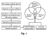
- FIG. 1 schematically illustrates one embodiment, of a method of selecting antibodies according to the present invention.
- the naive phage antibody library was first counterselected on a panel of non-tumorigenic cell lines to remove binders to common cell surface antigens (not shown) and then selected on live tumor cells under internalizing conditions to generate a sublibrary that is enriched for binders to internalizing cell surface epitopes. Further selection of this sublibrary on tissue slides by LCM enriched scFv fragments that bind to tumor cells in situ. Monoclonal phage antibodies were identified by screening selection output on tumor cell lines followed by rescreening positive clones on tissue slides. This selection scheme effectively restricts selection outcomes to phage antibodies that bind to epitopes present on both tumor cell lines and tumor cells in situ from actual cases. Moreover these antibodies are expected to possess internalizing functions that can be exploited for targeted payload delivery.
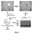
- FIG. 2 illustrates selection of phage antibody library on tissue slides by LCM.
- Tissue pieces containing tumor cells and tumor-bound phage were procured by Leica AS LMD and collected on the cap of a PCR tube (step 1 ).
- scFv-coding regions were amplified by PCR (step 2) and spliced into a phage display vector to create LCM secondary libraries (step 3) that were used for screening ( step 4) or additional rounds of selection ( step 5).
- Ctr control
- MW molecular weight.
- Figure 3 illustrates initial screening of selection output. FACS analysis was performed on tumor cell lines to identify positive clones, restricting the number of phage antibody that needed to be screened on tissue slides. Ctr, helper phage. Clones 1 ⁇ 4, four positive clones randomly chosen from the output following one round of LCM-based selection. Because these antibodies bound to both PC3 and Du-145 cells, it is likely that they bind to tumor antigens instead of artifacts associated with slide preparation. Tumor specificity and clinical relevance were further studied by IHC. PE-A, phycoerythrin channel; FITC-A, FITC channel.
- FIG. 4 panels A-C, show the results of immunohistochemistry studies. Biotinylated scFv fragments were used to stain CaP tissues.
- the UA20 scFv was originally isolated from selection on paraffin-embedded tissues; it stained tumor cells in both frozen and paraffin-embedded tissue slides.
- the 585II41 scFv was originally isolated from selection on frozen tissues; it stained tumor cells in frozen but not paraffin-embedded tissue slides.
- Panel A staining of frozen tissues with UA20 scFv.
- Panel B staining of frozen tissues with 585II41 scFv.
- Panel C staining of paraffin-embedded CaP tissues with UA20 scFv.
- Figures 5A, 5B , 5C illustrate the internalization of immunoliposomes. Fluorescent liposomes conjugated with the UA20 scFv were tested for internalization into prostate cancer cells.
- Figure 5A microscopic examination of uptake of UA20-ILs by PC3 and Du-145 cells. There was no uptake by BPH-1 cells.
- 5B FACS analysis of uptake of UA20-DiICl8(3)-DS-ILs by Du-145 cells. MFI, mean fluorescence intensity.
- Figure 5C quantification of UA20 scFv-IL uptake by prostate cancer and control cells. MFI values were obtained from FACS. NT-LPs, non-targeted liposomes.
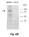
- Figures 6A and 6B illustrate the identification of ALCAM/MEMD/CD166 as the target of the 585II41 scFv.
- Figure 6A shows that binding of the 585II41 scFv to prostate cancer cells was specifically competed by a previously identified anti-ALCAM scFv, H3, and its corresponding IgG1 but not by a control scFv, OA12, and its corresponding IgG1.
- Figure 6B illustrates analysis of IP products by Western blot.
- Lysates from biotin surface-labeled Du-145 cells were incubated with 585II41 scFv and OA12 scFv (control) to generate IP products that were analyzed by Western blot using an ALCAM-specific commercial monoclonal antibody. Only the 585II41 scFv IP product reacted with the anti-ALCAM mAb.
- the band (indicated by an arrow) is located between 100 and 110 kDa.
- ALCAM is predicted to be a 65-kDa protein, but glycosylation causes it to appear as a band of ⁇ 105 kDa on SDS-PAGE, consistent with previous reports ( Saifullah et al. (2004) J. Immunol. 173: 6125-6133 ).
- MFI mean fluorescence intensity
- Figure 7 shows the amino acid sequences of internalizing prostate cancer specific antibodies: 3051.1 (SEQ ID NO:3), G12FC3 (SEQ ID NO:4), M6c42b (SEQ ID NO:5), 4F3YW (SEQ ID NO:6), M40pr146 (SEQ ID NO:7), UA20 (SEQ ID NO:8), UA8 (SEQ ID NO:9), 585II41 (SEQ ID ⁇ NO:10), 585II41.1 (SEQ ID NO:11), 585II56 (SEQ ID NO:12), 3076 (SEQ ID NO:13), and 3051 (SEQ ID NO:14).
- Figure 8 shows the amino acid sequences of internalizing prostate cancer specific antibodies: M49R (SEQ ID NO:15), RCI-14 (SEQ ID NO:16), II79_4 (SEQ ID NO:17), II79_3 (SEQ ID NO:18), T5II-4B.1 (SEQ ID NO:19), T5II-4B.2 (SEQ ID NO:20), RCI-11 (SEQ ID NO:21), RCI-20 (SEQ ID NO:22), CI-11A (SEQ ID NO:23), CI-14A (SEQ ID NO:24), and S95-2 (SEQ ID NO:25).
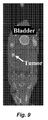
- Figure 9 shows the results of SPECT/CT imaging of UA20 scFv targeting to prostate cancer Du-145 xenograft tumor (arrow) in nude mice.
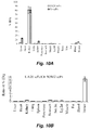
- Figures 10A, and 10B show the results of biodistribution studies.
- A SPECT/CT imaging of UA20 scFv targeting to prostate cancer Du-145 xenograft tumor (arrow) in nude mice.
- Figure 10B Biodistribution study. The values of %ID/g tissue for both the UA20 scFv and the control N3M2 scFv were plotted for tumor, blood and other organs/tissues. Standard errors are indicated. Sm.Int., small intestine. Lg.Int., large intestine.
- this invention pertains to a method that allows selection of phage antibodies against tumor cells in situ on fresh, fresh frozen, and paraffin-embedded tissues using laser capture microdissection.
- Laser capture microdissection allows small clusters of homogenous cells to be isolated and removed from tissue sections under direct microscopic visualization.
- the methods involve providing a display library (e.g ., a yeast- or phage-display library), contacting a tissue compromising the target type with members of the library; isolating groups of cells from the tissue using laser capture microdissection; and recovering members of the library that bind to cells in the isolated groups.
- a display library e.g ., a yeast- or phage-display library
- libraries can be used to screen for markers expressed in situ on essentially any desired cell type.
- binders e.g., antibodies
- binders specific for any pathological cell type, where the pathological cell displays different markers, than other cells, can be identified.
- the display library can optionally be counter selected on cells for which binding is not desired (e.g., normal healthy cells) and/or pre-selected, e.g., on a panel of target cells to enhance the representation of binders and/or internalizing members in the library.
- the library is created by selecting, e.g ., a naive phage antibody display library on a panel of target cells (e.g., tumor cell lines), and where internalization is desired, under internalizing conditions. Methods of preparation and selection of a phage antibody display library have been described, for example by Liu et al. (2004) Cancer Res. 64: 704-710 , and Poul et al. (2000) J. Mol. Biol.
- the phage library is preincubated with a panel of non-tumorigenic cells including, for example BPH-1, human mammary epithelial cells, MCF10A, and human fibroblasts to remove binders to common cell surface antigens.
- the predepleted library can then, optionally be incubated with a panel of target cells (e.g., prostate cancer cell lines (PC3 and Du-145)) at 37°C for 2 h; washed twice with 100 mM glycine, pH 2.8, in the presence of 150 mM NaCl; and washed once with PBS, pH 7.0.
- target cells e.g., prostate cancer cell lines (PC3 and Du-145)
- Internalized phage can be recovered by lysing the cells with 100 mM triethylamine, propagated in TG1, and purified by precipitation with polyethylene glycol 8000 as described previously (1), thereby creating a sublibrary that is enriched for binders to internalizing cell surface molecules.
- the library can then be incubated (selected against) one or more tissues containing the target cells.
- selections can be performed, on fresh, frozen, and/or paraffin embedded tissues.
- sections of the target tissue can be placed on microscope slides, and incubated with the library, for 1/2 to several hours, for example, at room temperature (e.g., 1 hour at room temperature).
- the tissue is then washed to remove unbound library members and prepared for laser capture microdissection according to standard methods.
- LCM laser capture microdissetion
- 5-500, more typically 100 or 200, still more typically 20-100 or 20-50 cells are procured at a time, e.g ., by generating a closed laser path around the group of cells of interest.
- the cells can then be collected (e.g., dropped into collection tubes by electrostatic force and gravity), and the bound and/or internalized library members recovered.
- phage bound to LCM-procured tissue pieces appear to lose the ability to infect bacteria, thereby posing a challenge to library selection. Little bacterial growth was observed under various culture conditions. This phenomenon was seen even in manually dissected tissue pieces that were not exposed to the UV laser used in the Leica LMD system. Exposure to ethanol during slide preparation for LCM seems to be a factor contributing to the observed reduction in phage viability.
- the problem is circumvented by using the genomes of phages (or yeast) bound to the procured cancer cell pieces as templates for amplification of scFv genes, e.g ., by PCR.
- the amplified scFv genes can easily be identified and/or sequenced.
- this invention provides a number of antibodies that specifically bind and are internalized into human prostate cancer cells.
- the antibodies were identified by selecting human antibody gene diversity libraries directly on the surface of prostate cancer cells in vivo using laser microdissection methods as described above and in the examples. Antibodies were identified that specifically bind and enter prostate cancer cells, with little or no binding to control cells.
- the antibodies in the library were expressed as single chain Fv (scFv) antibodies comprising a variable heavy (V H ) region linked to a variable light (V L ) region by a peptide linker, although it will be recognized that using the antibody sequence presented herein other forms of the antibodies can be provided.
- scFv single chain Fv
- V H and V L domains are illustrated in Tables 1 and 2, respectively as well as in Figures 7 and 8 .
- VH variable heavy chain of prostate cancer specific internalizing antibodies.
- Heavy chain Clone Frame 1 CDR1 Frame 2
- CDR2 Frame 3 CDR3 Frame 4 3051.1 SYGMY (SEQ ID NO:27 ) G12FC3 GSGMH (SEQ ID NO:34 ) M6c42b TYAMR (SEQ ID NO:41 ) 4F3YW SYAMH (SEQ ID NO:48 ) M40pr146 SYAMS (SEQ ID NO:55 ) UA20 NAWMN (SEQ ID NO:62 ) UA8 SFGMH (SEQ ID NO:69 ) 585II41 SYAMG (SEQ ID NO:76 ) 585II41.1 SYAMS (SEQ ID NO:83 ) 585II56 SYAMS (SEQ ID NO:90 ) 3076 GYWMS (SEQ ID NO:97 ) 3051 SYGMY (SEQ ID NO:10 4) M49R DHYMD (SEQ ID NO:11 1) R
- VL variable light chain of prostate cancer specific internalizing antibodies.
- Light Chain Clone Frame 1 CDR1 Frame 2
- CDR2 Frame 3 CDR3 Frame 4 3051.1 G12FC3 M6c42b 4F3YW M40pr146 UA20 UA8 585II41 585II41.1 585II56 3076 3051 M49R RCI-14 II79_4 II79_3 T5II ⁇ 4B.1 T5II ⁇ 4B.2 RCI ⁇ 11 RCI-20 CI-11A CI-14A S95-2
- variable heavy (VH) region is coupled to the variable light (V L ) either directly, or more preferably by a peptide linker (e.g., (Gly 4 Ser) 3 , SEQ ID NO:1).
- a peptide linker e.g., (Gly 4 Ser) 3 , SEQ ID NO:1.
- Illustrative scFv antibodies are shown in Table 3.
- the antibodies 3051.1, G12FC3, M6c42b, 4F3YW, M40pr146, UA20, UA8, 585II41, 585II41.1, 585II56, 3076, 3051, M49R, RCI-14, II79_4, II79_3, T511-4B.1, T5II-4B.2, RCI-11, RCI-20, CI-11A, CI-14A, and S95-2, or antibodies comprising one or more of the CDRs comprising these antibodies, or antibodies comprising the VH and/or VL domain(s) of these antibodies can readily be prepared using standard methods (e.g. chemical synthesis methods and/or recombinant expression methods) well known to those of skill in the art.
- prostate cancer specific antibodies can be identified by screening for antibodies that bind to the same epitope (e.g. that compete with the listed antibodies for binding to a prostate cancer cell) and/or by modification of the antibodies identified herein (e.g., 3051.1, G12FC3, M6c42b, 4F3YW, M40pr146, UA20, UA8, 585II41, 585II41.1, 585II56, 3076, 3051, M49R, RCI-14, II79_4, II79_3, T5II-4B.1, T5II-4B.2, RCI-11, RCI-20, CI-11A, CI-14A, and/or S95-2) to produce libraries of modified antibody and then rescreening antibodies in the library for improved binding to prostate cancer cells, and/or by screening of various libraries on prostate cancer cells, e.g ., as illustrated in Example 1 .
- the prostate cancer specific antibodies of this invention e.g., 3051.1, G12FC3, M6c42b, 4F3YW, M40pr146, UA20, UA8, 585II41, 585II41.1, 585II56, 3076, 3051, M49R, RCI-14, II79_4, II79_3, T511-4B.1, T5II-4B.2, RCI-11, RCI-20, CI-11A, CI-14A, S95-2, etc. ) , or variants thereof, can be chemically synthesized using well known methods of peptide synthesis.
- Solid phase synthesis in which the C-terminal amino acid of the sequence is attached to an insoluble support followed by sequential addition of the remaining amino acids in the sequence is one preferred method for the chemical synthesis of single chain antibodies.
- Techniques for solid phase synthesis are described by Barany and Merrifield, Solid Phase Peptide Synthesis; pp. 3-284 in The Peptides: Analysis, Synthesis, Biology. Vol. 2: Special Methods in Peptide Synthesis, Part A ., Merrifield et al. (1963) J. Am. Chem. Soc., 85: 2149-2156 , and Stewart et al. (1984) Solid Phase Peptide Synthesis, 2nd ed. Pierce Chem. Co., Rockford, I11 .
- the prostate cancer specific antibodies of this invention e.g., 3051.1, G12FC3, M6c42b, 4F3YW, M40pr146, UA20, UA8, 585II41, 585II41.1, 585II56, 3076, 3051, M49R, RCI-14, II79_4, II79_3, T5II-4B.1, T5II-4B.2, RCI-11, RCI-20, CI-11A, CI-14A, S95-2, etc. ) , or variants thereof, are prepared using standard techniques well known to those of skill in the art.
- nucleic acids encoding the desired antibody can be chemically synthesized according to a number of standard methods known to those of skill in the art. Oligonucleotide synthesis, is preferably carried out on commercially available solid phase oligonucleotide synthesis machines ( Needham-VanDevanter et al. (1984) Nucleic Acids Res. 12: 6159-6168 ) or manually synthesized using the solid phase phosphoramidite triester method described by Beaucage et. al. ( Beaucage et. al. (1981) Tetrahedron Letts. 22(20): 1859-1862 ). Alternatively, nucleic acids encoding the antibody can be amplified and/or cloned according to standard methods.
- prostate cancer specific internalizing antibodies e.g., 3051.1, G12FC3, M6c42b, 4F3YW, M40pr146, UA20, UA8, 585II41, 585II41.1, 585II56, 3076, 3051, M49R, RCI-14, II79_4, II79_3, T5II-4B.1, T5II-4B.2, RCI-11, RCI-20, CI-11A, CI-14A, S95-2), other "related" internalizing prostate cancer specific antibodies can be identified by screening for antibodies that cross-react with the identified antibodies, either at the epitope bound by the antibodies, and/or for antibodies that cross- react with the identified antibodies for binding to a prostate cancer cell (e.g., CaP cells, PC3 cells, etc.
- a prostate cancer cell e.g., CaP cells, PC3 cells, etc.
- the idiotype represents the highly variable antigen-binding site of an antibody and is itself immunogenic. During the generation of an antibody-mediated immune response, an individual will develop antibodies to the antigen as well as anti-idiotype antibodies, whose immunogenic binding site (idiotype) mimics the antigen.
- Anti-idiotypic antibodies can be raised against the variable regions of the antibodies identified herein using standard methods well known to those of skill in the art. Briefly, anti-idiotype antibodies can be made by injecting the antibodies of this invention, or fragments thereof (e.g., CDRs) into an animal thereby eliciting antisera against various antigenic determinants on the antibody, including determinants in the idiotypic region.
- CDRs CDRs
- Anti-analyte antibodies are well known in the art. Large molecular weight antigens (greater than approx. 5000 Daltons) can be injected directly into animals, whereas small molecular weight compounds (less than approx. 5000 Daltons) are preferably coupled to a high molecular weight immunogenic carrier, usually a protein, to render them immunogenic.
- the antibodies produced in response to immunization can be utilized as serum, ascites fluid, an immunoglobulin (Ig) fraction, an IgG fraction, or as affinity-purified monospecific material.
- Polyclonal anti-idiotype antibodies can be prepared by immunizing an animal with the antibodies of this invention prepared as described above. In general, it is desirable to immunize an animal which is species and allotype-matched with the animal from which the antibody (e.g. phage-display library) was derived. This minimizes the production of antibodies directed against non-idiotypic determinants.
- the antiserum so obtained is then usually absorbed extensively against normal serum from the same species from which the phage-display library was derived, thereby eliminating antibodies directed against non-idiotypic determinants. Absorption can be accomplished by passing antiserum over a gel formed by crosslinking normal (nonimmune) serum proteins with glutaraldehyde.
- Antibodies with anti-idiotypic specificity will pass directly through the gel, while those having specificity for non-idiotypic determinants will bind to the gel.
- Immobilizing nonimmune serum proteins on an insoluble polysaccharide support e.g., sepharose also provides a suitable matrix for absorption.
- Monoclonal anti-idiotype antibodies can be produced using the method of Kohler et al. (1975) Nature 256: 495 .
- monoclonal anti-idiotype antibodies can be prepared using hybridoma technology which comprises fusing (1)spleen cells from a mouse immunized with the antigen or hapten-carrier conjugate of interest (i.e., the antibodies or this invention or subsequences thereof) to (2) a mouse myeloma cell line which has been selected for resistance to a drug (e.g., 8-azaguanine).
- a myeloma cell line which does not secrete an immunoglobulin.
- Several such lines are known in the art.
- One generally preferred cell line is P3X63Ag8.653. This cell line is on deposit at the American Type Culture Collection as CRL-1580.
- Fusion can be carried out in the presence of polyethylene glycol according to established methods (see, e.g., Monoclonal Antibodies, R. Kennett, J. McKearn & K. Bechtol, eds. N.Y., Plenum Press, 1980 , and Current Topics in Microbiology & Immunology, Vol. 81, F. Melchers, M. Potter & N. L. Warner, eds., N.Y., Springer-Verlag, 1978 ).
- the resultant mixture of fused and unfused cells is plated out in hypoxanthine-aminopterin-thymidine (HAT) selective medium. Under these conditions, only hybrid cells will grow.
- HAT hypoxanthine-aminopterin-thymidine
- the culture medium is harvested and screened for the presence of monoclonal idiotypic, anti-analyte antibody by any one of a number of methods which include solid phase RIA and enzyme-linked immunosorbent assay.
- Cells from culture wells containing antibody of the desired specificity are then expanded and recloned. Cells from those cultures that remain positive for the antibody of interest are then usually passed as ascites tumors in susceptible, histocompatible, pristane-primed mice.
- Ascites fluid is harvested by tapping the peritoneal cavity, retested for antibody, and purified as described above. If a nonsecreting myeloma line is used in the fusion, affinity purification of the monoclonal antibody is not usually necessary since the antibody is already homogeneous with respect to its antigen-binding characteristics. All that is necessary is to isolate it from contaminating proteins in ascites, i.e., to produce an immunoglobulin fraction.
- the hybrid cell lines of interest can be grown in serum-free tissue culture and the antibody harvested from the culture medium. In general, this is a less desirable method of obtaining large quantities of antibody because the yield is low. It is also possible to pass the cells intravenously in mice and to harvest the antibody from serum. This method is generally not preferred because of the small quantity of serum which can be obtained per bleed and because of the need for extensive purification from other serum components. However, some hybridomas will not grow as ascites tumors and therefore one of these alternative methods of obtaining antibody must be used.
- prostate cancer specific antibodies of this invention can be identified by the fact that they bind the same epitope as the "prototypic" antibodies of this invention (e.g 3051.1, G12FC3, M6c42b, 4F3YW, M40pr146, UA20, UA8, 585II41, 585II41.1, 585II56, 3076, 3051, M49R, RCI-14, II79_4, II79_3, T5II-4B.1, T5II-4B.2, RCI-11, RCI-20, CI-11A, CI-14A, S95-2, etc. ) .
- To identify such antibodies it s not necessary to isolate the subject epitope.
- a prostate cancer cell e.g. a CaP cell, a PC3 cell, etc.
- epitope bound by the prototypic antibodies of this invention is determined e.g. by epitope mapping techniques. Methods of epitope mapping are well known to those of skill in the art (see, e.g., Reyes et al. (1992) Hepatitis E Virus (HEV): Epitope Mapping and Detection of Strain Variation, Elsevier Science Publisher Shikata et al. eds., Chapter 43:237-245 ; Li et al. (1993) Nature 363: 85-88 ). Epitope mapping can be performed using Novatope system, a kit for which is commercially available from Novagen, Inc.
- Novatope system a kit for which is commercially available from Novagen, Inc.
- cross-reactive prostate cancer specific antibodies show at least 60%, preferably 80%, more preferably 90%, and most preferably at least 95% or at least 99% cross-reactivity with one or more of the prototypic antibodies of this invention.
- a mutant scFv gene repertoire can be created containing a V H gene of the prototypic antibodies (e.g. as shown in Tables 1-3, and/or Figures 7 and 8 ) antibody and a human V L gene repertoire (light chain shuffling).
- the scFv gene repertoire can be cloned into a phage display vector, e.g., pHEN-1 ( Hoogenboom et al. (1991) Nucleic Acids Res., 19: 4133-4137 ) or other vectors, e.g. as described herein in the examples, and after transformation a library of transformants is obtained.
- the prostate cancer specific antibody e.g., 3051.1, G12FC3, M6c42b, 4F3YW, M40pr146, UA20, UA8, 585II41, 585II41.1, 585II56, 3076, 3051, M49R, RCI-14, II79_4, II79_3, T5II-4B.1, T5II-4B.2, RCI-11, RCI-20, CI-11A, CI-14A, S95-2, etc.
- the prostate cancer specific antibody e.g., 3051.1, G12FC3, M6c42b, 4F3YW, M40pr146, UA20, UA8, 585II41, 585II41.1, 585II56, 3076, 3051, M49R, RCI-14, II79_4, II79_3, T5II-4B.1, T5II-4B.2, RCI-11, RCI-20, CI-11A, CI-14A, S95-2, etc.
- V H CDR1 and/or CDR2, and/or CDR3 and light chain are cloned into a vector containing a human V H gene repertoire to create a phage antibody library transformants.
- chain shuffling to increase antibody affinity see Schier et al. (1996) J. Mol. Biol., 255: 28-43 , and the like.
- CDRs complementarity determining regions
- V H V H
- V L V L
- CDR1, CDR2, and CDR3 V L
- mutant antibody libraries can be created where partial or entire CDRs are randomized (V L CDR1 CDR2 and/or CDR3 and/or V H CDR1, CDR2 and/or CDR3).
- each CDR is randomized in a separate library, using a known antibody (e.g., 3051.1, G12FC3, M6c42b, 4F3YW, M40pr146, UA20, UA8, 585II41, 585II41.1, 585II56, 3076, 3051, M49R, RCI-14, II79_4, II79_3, T5II-4B.1, T5II-4B.2, RCI-11, RCI-20, CI-11A, CI-14A, and/or S95-2) as a template.
- a known antibody e.g., 3051.1, G12FC3, M6c42b, 4F3YW, M40pr146, UA20, UA8, 585II41, 585II41.1, 585
- V H CDR3 often occupies the center of the binding pocket, and thus mutations in this region are likely to result in an increase in affinity ( Clackson et al. (1995) Science, 267: 383-386 ).
- four V H CDR3 residues are randomized at a time using the nucleotides NNS ( see, e.g., Schier et al. (1996) Gene, 169: 147-155 ; Schier and Marks (1996) Human Antibodies and Hybridomas. 7: 97-105, 1996 ; and Schier et al. (1996) J. Mol. Biol. 263: 551-567 ).
- antibody forms include, but are not limited to multivalent antibodies, full antibodies, scFv, (scFv') 2 , Fab, (Fab') 2 , chimeric antibodies, and the like.
- two prostate cancer specific scFvs are joined, either through a linker (e.g., a carbon linker, a peptide, etc.) or through a disulfide bond between, for example, two cysteins.
- a linker e.g., a carbon linker, a peptide, etc.
- a disulfide bond between, for example, two cysteins.
- a cysteine residue can be introduced by site directed mutagenesis at the carboxy-terminus of the antibodies described herein.
- scFv can be expressed from this construct, purified by IMAC, and analyzed by gel filtration.
- cysteine is reduced by incubation with 1 mM 3-mercaptoethanol, and half of the scFv blocked by the addition of DTNB. Blocked and unblocked scFvs are incubated together to form (scFv') 2 and the resulting material can be analyzed by gel filtration.
- the affinity of the resulting dimmer can be determined using standard methods, e.g . by BIAcore.
- the (scFv') 2 dimer is created by joining the scFv' fragments through a linker, more preferably through a peptide linker.
- a linker more preferably through a peptide linker.
- This can be accomplished by a wide variety of means well known to those of skill in the art. For example, one preferred approach is described by Holliger et al. (1993) Proc. Natl. Acad. Sci. USA, 90: 6444-6448 ( see also WO 94/13804 ).
- Fabs and (Fab') 2 dimers can also readily be prepared.
- Fab is a light chain joined to V H -C H 1 by a disulfide bond and can readily be created using standard methods known to those of skill in the art.
- the F(ab)' 2 can be produced by dimerizing the Fab, e.g. as described above for the (scFv') 2 dimer.
- the antibodies of this invention also include "chimeric" antibodies in which a portion of the heavy and/or light chain is identical with or homologous to corresponding sequences in antibodies derived from a particular species or belonging to a particular antibody class or subclass, while the remainder of the chain(s) is identical with or homologous to corresponding sequences in antibodies derived from another species or belonging to another antibody class or subclass, as well as fragments of such antibodies, so long as they exhibit the desired biological activity ( see, e.g., U.S. Pat. No. 4,816,567 ; Morrison et al. (1984) Proc. Natl. Acad. Sci. 81: 6851-6855 , etc. ).
- Chimeric antibodies are antibodies comprising a portions from two different species (e.g. a human and non-human portion).
- the antigen combining region (or variable region) of a chimeric antibody is derived from a one species source and the constant region of the chimeric antibody (which confers biological effector function to the immunoglobulin) is derived from another source.
- a large number of methods of generating chimeric antibodies are well known to those of skill in the art (see, e.g., U.S.
- the procedures used to produce chimeric antibodies consist of the following steps (the order of some steps may be interchanged): (a) identifying and cloning the correct gene segment encoding the antigen binding portion of the antibody molecule; this gene segment (known as the VDJ, variable, diversity and joining regions for heavy chains or VJ, variable, joining regions for light chains, or simply as the V or variable region or V H and V L regions) may be in either the cDNA or genomic form; (b) cloning the gene segments encoding the human constant region or desired part thereof; (c) ligating the variable region to the constant region so that the complete chimeric antibody is encoded in a transcribable and translatable form; (d) ligating this construct into a vector containing a selectable marker and gene control regions such as promoters, enhancers and poly(A) addition signals; (e) amplifying this construct in a host cell (e.g ., bacteria); (f) introducing the DNA into eukaryotic cells (trans
- Antibodies of several distinct antigen binding specificities have been manipulated by these protocols to produce chimeric proteins (e.g ., anti-TNP: Boulianne et al. (1984) Nature, 312: 643 ; and anti-tumor antigens: Sahagan et al. (1986) J. Immunol., 137: 1066 ).
- anti-TNP Boulianne et al. (1984) Nature, 312: 643
- anti-tumor antigens Sahagan et al. (1986) J. Immunol., 137: 1066
- effector functions have been achieved by linking new sequences to those encoding the antigen binding region. Some of these include enzymes ( Neuberger et al. (1984) Nature 312: 604 ), immunoglobulin constant regions from another species and constant regions of another immunoglobulin chain ( Sharon et al. (1984) Nature 309: 364 ; Tan et al., (1985) J. Immunol. 135
- a recombinant DNA vector is used to transfect a cell line that produces a prostate cancer specific antibody of this invention.
- the novel recombinant DNA vector contains a "replacement gene" to replace all or a portion of the gene encoding the immunoglobulin constant region in the cell line (e.g., a replacement gene may encode all or a portion of a constant region of a human immunoglobulin, a specific immunoglobulin class, or an enzyme, a toxin, a biologically active peptide, a growth factor, inhibitor, or a linker peptide to facilitate conjugation to a drug, toxin, or other molecule, etc.), and a "target sequence” that allows for targeted homologous recombination with immunoglobulin sequences within the antibody producing cell.
- a recombinant DNA vector is used to transfect a cell line that produces an antibody having a desired effector function, (e.g ., a constant region of a human immunoglobulin) in which case, the replacement gene contained in the recombinant vector may encode all or a portion of a region of a prostate cancer specific antibody of this invention and the target sequence contained in the recombinant vector allows for homologous recombination and targeted gene modification within the antibody producing cell.
- a desired effector function e.g ., a constant region of a human immunoglobulin
- the resulting chimeric antibody when only a portion of the variable or constant region is replaced, can define the same antigen and/or have the same effector function yet be altered or improved so that the chimeric antibody may demonstrate a greater antigen specificity, greater affinity binding constant, increased effector function, or increased secretion and production by the transfected antibody producing cell line, etc.
- the processes of selection for integrated DNA (via a selectable marker), screening for chimeric antibody production, and cell cloning can be used to obtain a clone of cells producing the chimeric antibody.
- a piece of DNA that encodes a modification for a monoclonal antibody can be targeted directly to the site of the expressed immunoglobulin gene within a B-cell or hybridoma cell line.
- DNA constructs for any particular modification can be made to alter the protein product of any monoclonal cell line or hybridoma.
- the level of expression of chimeric antibody should be higher when the gene is at its natural chromosomal location rather than at a random position.
- Detailed methods for preparation of chimeric (humanized) antibodies can be found in U.S. Patent 5,482,856 .
- this invention provides for intact, fully human prostate cancer specific antibodies.
- Such antibodies can readily be produced in a manner analogous to making chimeric human antibodies.
- the fully human recognition function e.g., VH and V L
- VH and V L the fully human recognition function of the antibodies described herein is utilized.
- this invention contemplates diabodies comprising one or more of the V H and V L domains described herein.
- the term "diabodies” refers to antibody fragments typically having two antigen-binding sites.
- the fragments typically comprise a heavy chain variable domain (V H ) connected to a light chain variable domain (V L ) in the same polypeptide chain (V H -V L ).
- V H heavy chain variable domain
- V L light chain variable domain
- the domains are forced to pair with the complementary domains of another chain and create two antigen-binding sites.
- Diabodies are described more fully in, for example, EP 404,097 ; WO 93/11161 , and Holliger et al. (1993) Proc. Natl. Acad. Sci. USA 90: 6444-6448 .
- the antibodies of this invention can be constructed as unibodies.
- UniBody are antibody technology that produces a stable, smaller antibody format with an anticipated longer therapeutic window than certain small antibody formats.
- unibodies are produced from IgG4 antibodies by eliminating the hinge region of the antibody. Unlike the full size IgG4 antibody, the half molecule fragment is very stable and is termed a uniBody. Halving the IgG4 molecule leaves only one area on the UniBody that can bind to a target. Methods of producing unibodies are described in detail in PCT Publication WO2007/059782 , which is incorporated herein by reference in its entirety (see, also, Kolfschoten et al. (2007) Science 317: 1554-1557 ).
- sequence information provided herein is used to construct affibody molecules that bind prostate cancer cells.
- Affibody molecules are class of affinity proteins based on a 58-amino acid residue protein domain, derived from one of the IgG-binding domains of staphylococcal protein A. This three helix bundle domain has been used as a scaffold for the construction of combinatorial phagemid libraries, from which affibody variants that target the desired molecules can be selected using phage display technology ( see, e.g,. Nord et al. (1997) Nat. Biotechnol. 15: 772-777 ; Ronmark et al. (2002) Eur. J. Biochem., 269: 2647-2655 .). Details of Affibodies and methods of production are known to those of skill (see, e.g., US Patent No 5,831,012 which is incorporated herein by reference in its entirety).
- the antibodies described above can be provided as whole intact antibodies (e.g ., IgG), antibody fragments, or single chain antibodies, using methods well known to those of skill in the art.
- the antibody can be from essentially any mammalian species, to reduce immunogenicity, it is desirable to use an antibody that is of the species in which the antibody and/or chimeric moiety is to be used. In other words, for use in a human, it is desirable to use a human, humanized, or chimeric human antibody.
- selection for increased avidity can involves measuring the affinity of the antibody for the target antigen (e.g ., a prostate cancer cell).
- the K d of the antibody is determined from the kinetics of binding to, e.g . the target cell in a BIAcore, a biosensor based on surface plasmon resonance.
- the antigen or cell is coupled to a derivatized sensor chip capable of detecting changes in mass. When antibody is passed over the sensor chip, antibody binds to the antigen resulting in an increase in mass that is quantifiable.
- Measurement of the rate of association as a function of antibody concentration can be used to calculate the association rate constant (k on ).
- buffer is passed over the chip and the rate of dissociation of antibody (k off ) determined.
- K on is typically measured in the range 1.0 x 10 2 to 5.0 x 10 6 and k off in the range 1.0 x 10 -1 to 1.0 x 10 -6 .
- the equilibrium constant K d is often calculated as k off /k on and thus is typically measured in the range 10 -5 to 10 -12 . Affinities measured in this manner correlate well with affinities measured in solution by fluorescence quench titration.
- the prototypical prostate cancer -specific antibodies of this invention e.g., 3051.1, G12FC3, M6c42b, 4F3YW, M40pr146, UA20, UA8, 585II41, 585II41.1, 585II56, 3076, 3051, M49R, RCI-14,II79_4, II79_3, T5II-4B.1, T5II-4B.2, RCI-11, RCI-20, CI-11A, CI-14A, S95-2, etc. ) specifically bind to and are internalized by prostate cancer cells.
- the antibodies can be used alone as therapeutics ( e.g .
- an effector e.g . an effector molecule such as a cytotoxin, a radiolabel, a cancer drug, etc.
- various prostate cancer cells e.g . isolated cells, metastatic cells, solid tumor cells, etc.
- the antibodies of this invention are utilized in a "pretargeting" strategy (resulting in formation of a chimeric moiety at the target site after administration of the effector moiety) or in a “targeting” strategy where the antibody is coupled to an effector molecule prior to use to provide a chimeric molecule.
- a chimeric molecule or chimeric composition or chimeric moiety refers to a molecule or composition wherein two or more molecules that exist separately in their native state are joined together to form a single molecule having the desired functionality of its constituent molecules.
- one of the constituent molecules of a chimeric molecule is a "targeting molecule.
- the targeting molecule is a molecule such as a ligand or an antibody that specifically binds (and, in certain embodiments, is internalized) by its corresponding target, e.g ., a prostate cancer cell.
- effector refers to a molecule or group of molecules that is to be specifically/preferentially transported toor into the target cell (e.g ., a prostate cancer cell). It is noted that in this context, such specific transport need not be exclusively to or into a cancer cell, but merely need to provide preferential delivery of the effector to or into the cancer cell as compared to normal healthy cells.
- the effector molecule typically has a characteristic activity that is to be delivered to or into the target cell.
- Effector molecules include, but are not limited to cytotoxins, labels, radionuclides, ligands, antibodies, drugs, liposomes, nanoparticles, viral particles, cytokines, and the like.
- the effector comprises a detectable label.
- Suitable detectable labels include, but are not limited to radio-opaque labels, nanoparticles, PET labels, MRI labels, radioactive labels, and the like.
- radionuclides and useful in various embodiments of the present invention gamma-emitters, positron-emitters, x-ray emitters and fluorescence-emitters are suitable for localization, diagnosis and/or staging, and/or therapy, while beta and alpha-emitters and electron and neutron-capturing agents, such as boron and uranium, also can be used for therapy.
- the detectable labels can be used in conjunction with an external detector and/or an internal detector and provide a means of effectively localizing and/or visualizing prostate cancer cells. Such detection/visualization can be useful in various contexts including, but not limited to pre-operative and intraoperative settings.
- this invention relates to a method of intraoperatively detecting and prostate cancers in the body of a mammal. These methods typically involve administering to the mammal a composition comprising, in a quantity sufficient for detection by a detector (e.g . a gamma detecting probe), an prostate cancer specific antibody labeled with a detectable label (e.g . antibodies of this invention labeled with a radioisotope, e.g.
- the label-bound antibody can be used in the technique of radioguided surgery, wherein relevant tissues in the body of a subject can be detected and located intraoperatively by means of a detector, e.g . a gamma detecting probe.
- a detector e.g . a gamma detecting probe.
- the surgeon can, intraoperatively, use this probe to find the tissues in which uptake of the compound labeled with a radioisotope, that is, e.g . a low-energy gamma photon emitter, has taken place.
- a radioisotope that is, e.g . a low-energy gamma photon emitter
- certain preferred effectors include, but are not limited to cytotoxins (e.g. Pseudomonas exotoxin, ricin, abrin, Diphtheria toxin, and the like), or cytotoxic drugs or prodrugs, in which case the chimeric molecule may act as a potent cell-killing agent specifically targeting the cytotoxin to prostate cancer cells.
- cytotoxins e.g. Pseudomonas exotoxin, ricin, abrin, Diphtheria toxin, and the like
- cytotoxic drugs or prodrugs in which case the chimeric molecule may act as a potent cell-killing agent specifically targeting the cytotoxin to prostate cancer cells.
- the effector can include a liposome encapsulating a drug (e.g . an anti-cancer drug such as abraxane, doxorubicin, pamidronate disodium, anastrozole, exemestane, cyclophosphamide, epirubicin, toremifene, letrozole, trastuzumab, megestroltamoxifen, paclitaxel, docetaxel, capecitabine, goserelin acetate, zoledronic acid, vinblastine, etc.
- a drug e.g . an anti-cancer drug such as abraxane, doxorubicin, pamidronate disodium, anastrozole, exemestane, cyclophosphamide, epirubicin, toremifene, letrozole, trastuzumab, megestroltamoxifen, paclitaxel, docetaxel, capecitabine,
- the chimeric moieties of this invention can be used to direct detectable labels to a tumor site. This can facilitate tumor detection and/or localization. It can be effective for detecting primary tumors, or, in certain embodiments, secondary tumors produced by, e.g ., prostate metastatic cells.
- the effector component of the chimeric moiety comprises a "radio-opaque" label, e.g . a label that can be easily visualized using x-rays.
- Radio-opaque materials are well known to those of skill in the art. The most common radio-opaque materials include iodide, bromide or barium salts.
- radiopaque materials include, but are not limited to, organic bismuth derivatives (see, e.g., U.S. Patent 5,939,045 ), radio-opaque polyurethanes (see, e.g., U.S. Patent 5,346,981 ), organobismuth composites (see, e.g., U.S. Patent 5,256,334 ), radio-opaque barium polymer complexes (see, e.g., U.S. Patent 4,866,132 ), and the like.
- organic bismuth derivatives see, e.g., U.S. Patent 5,939,045
- radio-opaque polyurethanes see, e.g., U.S. Patent 5,346,981
- organobismuth composites see, e.g., U.S. Patent 5,256,334
- radio-opaque barium polymer complexes see, e.g., U.S. Patent 4,866,
- the antibodies of this invention can be coupled directly to the radio-opaque moiety or they can be attached to a "package" (e.g., a chelate, a liposome, a polymer microbead, a nanoparticle, etc .) carrying, containing, or comprising the radio-opaque material, e.g ., as described below.
- a "package” e.g., a chelate, a liposome, a polymer microbead, a nanoparticle, etc .
- Detectable labels suitable for use as the effector molecule component of the chimeric moietys of this invention include any composition detectable by spectroscopic, photochemical, biochemical, immunochemical, electrical, optical or chemical means.
- Useful labels in the present invention include magnetic beads (e.g.
- fluorescent dyes e.g ., fluorescein isothiocyanate, texas red, rhodamine, green fluorescent protein, and the like
- radiolabels e.g ., 3 H, 125 I, 35 S, 14 C, or 32 P
- enzymes e.g ., horse radish peroxidase, alkaline phosphatase and others commonly used in an ELISA
- colorimetric labels such as colloidal gold or colored glass or plastic ( e.g . polystyrene, polypropylene, latex, etc. ) beads, nanoparticles, quantum dots, and the like.
- suitable radiolabels include, but are not limited to, 99 Tc, 203 Pb, 67 Ga, 68 Ga, 72 As, 111 In, 113m In, 97 Ru, 62 Cu, 641 Cu, 52 Fe, 52m Mn, 51 Cr, 186 Re, 188 Re, 77 As, 90 Y, 67 Cu, 169 Er, 121 Sn, 127 Te, 142 Pr, 143 Pr, 198 Au, 199 Au, 161 Tb, 109 Pd, 165 Dy, 149 Pm, 151 Pm, 153 Sm, 157 Gd, 159 Gd, 166 Ho, 172 Tm, 169 Yb 175 Yb, 177 Lu, 105 Rh, and 111 Ag.
- Radiolabels may be detected using photographic film, scintillation detectors, PET imaging, MRI, and the like. Fluorescent markers can be detected using a photodetector to detect emitted illumination. Enzymatic labels are typically detected by providing the enzyme with a substrate and detecting the reaction product produced by the action of the enzyme on the substrate, and colorimetric labels are detected by simply visualizing the colored label.
- the effector can comprise a radiosensitizer that enhances the cytotoxic effect of ionizing radiation (e.g ., such as might be produced by 60 Co or an x-ray source) on a cell.
- a radiosensitizer that enhances the cytotoxic effect of ionizing radiation (e.g ., such as might be produced by 60 Co or an x-ray source) on a cell.
- radiosensitizing agents include, but are not limited to benzoporphyrin derivative compounds ( see, e.g., U.S. Patent 5,945,439 ), 1,2,4-benzotriazine oxides ( see, e.g., U.S. Patent 5,849,738 ), compounds containing certain diamines ( see, e.g., U.S. Patent 5,700,825 ), BCNT ( see, e.g., U.S.
- Patent 5,872,107 radiosensitizing nitrobenzoic acid amide derivatives (see, e.g., U.S. Patent 4,474,814 ), various heterocyclic derivatives (see, e.g., U.S. Patent 5,064,849 ), platinum complexes (see, e.g ., U.S. Patent 4,921,963 ), and the like.
- the effector can include an alpha emitter, i.e . a radioactive isotope that emits alpha particles.
- Alpha-emitters have recently been shown to be effective in the treatment of cancer ( see, e.g., McDevitt et al. (2001) Science 294:1537-1540 ; Ballangrud et al. (2001) Cancer Res. 61: 2008-2014 ; Borchardt et al. (2003) Cancer Res. 63: 5084-50 ).
- Suitable alpha emitters include, but are not limited to Bi, 213 Bi, 211 At, and the like.
- the effector molecule can also comprise a ligand, an epitope tag, or an antibody.
- preferred ligands and antibodies include those that bind to surface markers on immune cells. Chimeric moietys utilizing such antibodies as effector molecules act as bifunctional linkers establishing an association between the immune cells bearing binding partner for the ligand or antibody and the prostate cancer cell(s).
- the pharmaceuticals and/or radiolabels described herein can be provided as a chelate, particularly where a pre-targeting strategy is utilized.
- the chelating molecule is typically coupled to a molecule (e.g . biotin, avidin, streptavidin, etc. ) that specifically binds an epitope tag attached to a prostate cancer specific antibody of this invention.
- chelating groups are well known to those of skill in the art.
- chelating groups are derived from ethylene diamine tetra-acetic acid (EDTA), diethylene triamine penta-acetic acid (DTPA), cyclohexyl 1,2-diamine tetra-acetic acid (CDTA), ethyleneglycol-O,O'-bis(2-aminoethyl)-N,N,N',N'-tetra-acetic acid (EGTA), N,N-bis(hydroxybenzyl)-ethylenediamine-N,N'-diacetic acid (HBED), triethylene tetramine hexa-acetic acid (TTHA), 1,4,7,10-tetraazacyclododecane-N,N'-,N",N"'-tetra-acetic acid (DOTA), hydroxyethyldiamine triacetic acid (HEDTA), 1,4,8,11-tetra-azacyclotetradecane-N
- Examples of certain preferred chelators include unsubstituted or, substituted 2-iminothiolanes and 2-iminothiacyclohexanes, in particular 2-imino-4-mercaptomethylthiolane.
- chelating agent 1,4,7,10-tetraazacyclododecane-N, N, N", N"'-tetraacetic acid (DOTA)
- DOTA 1,4,7,10-tetraazacyclododecane-N, N, N", N"'-tetraacetic acid
- 5: 565-576 describes a similar method wherein one carboxyl group of DOTA is converted to an active ester, and the activated DOTA is mixed with an antibody, linking the antibody to DOTA via the epsilon-amino group of a lysine residue of the antibody, thereby converting one carboxyl group of DOTA to an amide moiety.
- the chelating agent can be coupled, directly or through a linker, to an epitope tag or to a moiety that binds an epitope tag.
- Conjugates of DOTA and biotin have been described (see, e.g., Su (1995) J. Nucl. Med., 36 (5 Suppl):154P , which discloses the linkage of DOTA to biotin via available amino side chain biotin derivatives such as DOTA-LC-biotin or DOTA-benzyl-4-(6-amino-caproamide)-biotin).
- Yau et al., WO 95/15335 disclose a method of producing nitro-benzyl-DOTA compounds that can be conjugated to biotin.
- the method comprises a cyclization reaction via transient projection of a hydroxy group; tosylation of an amine; deprotection of the transiently protected hydroxy group; tosylation of the deprotected hydroxy group; and intramolecular tosylate cyclization.
- Wu et al. (1992) Nucl. Med. Biol., 19(2): 239-244 discloses a synthesis of macrocylic chelating agents for radiolabeling proteins with 111 IN and 90 Y.
- Wu et al. makes a labeled DOTA-biotin conjugate to study the stability and biodistribution of conjugates with avidin, a model protein for studies. This conjugate was made using a biotin hydrazide which contained a free amino group to react with an in situ generated activated DOTA derivative.
- the antibodies of this invention can be used to deliver a variety of cytotoxic drugs including therapeutic drugs, a compound emitting radiation, molecules of plants, fungal, or bacterial origin, biological proteins, and mixtures thereof.
- the cytotoxic drugs can be intracellularly acting cytotoxic drugs, such as short-range radiation emitters, including, for example, short-range, high-energy ⁇ -emitters as described above.
- Enzymatically active toxins and fragments. thereof are exemplified by diphtheria toxin A fragment, nonbinding active fragments of diphtheria toxin, exotoxin A (from Pseudomonas aeruginosa ) , ricin A chain, abrin A chain, modeccin A chain, .alpha.-sacrin, certain Aleurites fordii proteins, certain Dianthin proteins, Phytolacca americana proteins (PAP, PAPII and PAP-S), Morodica charantia inhibitor, curcin, crotin, Saponaria officinalis inhibitor, gelonin, mitogillin, restrictocin, phenomycin, and enomycin, for example.
- the cytotoxins can include, but are not limited to Pseudomonas exotoxins, Diphtheria toxins, ricin, abrin and derivatives thereof.
- Pseudomonas exotoxin A is an extremely active monomeric protein (molecular weight 66 kD), secreted by Pseudomonas aeruginosa, which inhibits protein synthesis in eukaryotic cells through the inactivation of elongation factor 2 (EF-2) by catalyzing its ADP-ribosylation (catalyzing the transfer of the ADP ribosyl moiety of oxidized NAD onto EF-2).
- the toxin contains three structural domains that act in concert to cause cytotoxicity.
- Domain Ia (amino acids 1-252) mediates cell binding.
- Domain II (amino acids 253-364) is responsible for translocation into the cytosol and domain III (amino acids 400-613) mediates ADP ribosylation of elongation factor 2, which inactivates the protein and causes cell death.
- domain Ib (amino acids 365-399) remains undefined, although a large part of it, amino acids 365-380, can be deleted without loss of cytotoxicity. See Siegall et al. (1989) J. Biol. Chem. 264: 14256-14261 .
- the antibody is attached to a preferred molecule in which domain Ia (amino acids 1 through 252) is deleted and amino acids 365 to 380 have been deleted from domain Ib.
- domain Ia amino acids 1 through 252
- amino acids 365 to 380 have been deleted from domain Ib.
- all of domain Ib and a portion of domain II can be deleted, particularly if the deleted sequences are replaced with a linking peptide.
- PE and other cytotoxic proteins can be further modified using site-directed mutagenesis or other techniques known in the art, to alter the molecule for a particular desired application.
- site-directed mutagenesis or other techniques known in the art, to alter the molecule for a particular desired application.
- means to alter the PE molecule in a manner that does not substantially affect the functional advantages provided by the PE molecules described here can also be used and such resulting molecules are intended to be covered herein.
- diphtheria toxin kills cells by ADP-ribosylating elongation factor 2 thereby inhibiting protein synthesis. Diphtheria toxin, however, is divided into two chains, A and B, linked by a disulfide bridge. In contrast to PE, chain B of DT, which is on the carboxyl end, is responsible for receptor binding and chain A, which is present on the amino end, contains the enzymatic activity ( Uchida et al.(1972) Science, 175: 901-903 ; Uchida et al. (1973) J. Biol. Chem., 248: 3838-3844 ).
- the antibody-Diphtheria toxin chimeric moietys of this invention have the native receptor-binding domain removed by truncation of the Diphtheria toxin B chain.
- One illustrative modified Dipththeria toxin is DT388, a DT in which the carboxyl terminal sequence beginning at residue 389 is removed ( see, e.g., Chaudhary et al. (1991) Bioch. Biophys. Res. Comm., 180: 545-551 ).
- the DT molecules can be chemically conjugated to the prostate cancer specific antibody, but, in certain preferred embodiments, the antibody will be fused to the Diphtheria toxin by recombinant means (see, e.g., Williams et al. (1990) J. Biol. Chem. 265: 11885-11889 ).
- the effector comprises a viral particle (e.g ., a filamentous phage, an adeno-associated virus (AAV), a lentivirus, and the like).
- the antibody can be conjugated to the viral particle and/or can be expressed on the surface of the viral particle (e.g . a filamentous phage).
- the viral particle can additionally include a nucleic acid that is to be delivered to the target (e.g ., prostate cancer) cell.
- the use of viral particles to deliver nucleic acids to cells is described in detail in WO 99/55720 , US 6,670,188 , US 6,642,051 , and US 6,669,936 .
- Suitable effector molecules include pharmacological agents or encapsulation systems containing various pharmacological agents.
- the targeting molecule of the chimeric moiety can be attached directly to a drug that is to be delivered directly to the tumor.
- drugs are well known to those of skill in the art and include, but are not limited to, abraxane, doxorubicin, pamidronate disodium, anastrozole, exemestane, cyclophosphamide, epirubicin, toremifene, letrozole, trastuzumab, megestroltamoxifen, paclitaxel, docetaxel, capecitabine, goserelin acetate, zoledronic acid, vinblastine, etc. )" an antisense molecule, an SiRNA, and the like.
- the effector molecule can comprise an encapsulation system, such as a viral capsid, a liposome, or micelle that contains a therapeutic composition such as a drug, a nucleic acid (e.g . an antisense nucleic acid or another nucleic acid to be delivered to the cell), or another therapeutic moiety that is preferably shielded from direct exposure to the circulatory system.
- a nucleic acid e.g . an antisense nucleic acid or another nucleic acid to be delivered to the cell
- another therapeutic moiety that is preferably shielded from direct exposure to the circulatory system.
- Means of preparing liposomes attached to antibodies are well known to those of skill in the art. See, for example, U.S. Patent No. 4,957,735 , Connor et al. (1985) Pharm. Ther., 28: 341-365 .
- coupling of liposomes to antibodies of this invention is illustrated herein in the Examples.
- the antibodies of this invention and the effector molecule(s) can be joined together in any order.
- the effector molecule can be joined to either the amino or carboxy termini of the targeting molecule.
- the targeting molecule can also be joined to an internal region of the effector molecule, or conversely, the effector molecule can be joined to an internal location of the targeting molecule, as long as the attachment does not interfere with the respective activities of the molecules.
- the antibody and the effector can be attached by any of a number of means well known to those of skill in the art.
- the effector is conjugated, either directly or through a linker (spacer), to the targeting molecule.
- linker spacer
- both the effector molecule and the antibody are polypeptides it is preferable to recombinantly express the chimeric molecule as a single-chain fusion protein.
- the prostate cancer specific antibody is chemically conjugated to the effector molecule (e.g ., a cytotoxin, a label, a ligand, or a drug or liposome, etc. ).
- the effector molecule e.g ., a cytotoxin, a label, a ligand, or a drug or liposome, etc.
- Means of chemically conjugating molecules are well known to those of skill.
- Polypeptides typically contain variety of functional groups; e.g ., carboxylic acid (COOH) or free amine (-NH 2 ) groups, that are available for reaction with a suitable functional group on an effector molecule to bind the effector thereto.
- functional groups e.g ., carboxylic acid (COOH) or free amine (-NH 2 ) groups, that are available for reaction with a suitable functional group on an effector molecule to bind the effector thereto.
- the antibody and/or the effector can be derivatized to expose or attach additional reactive functional groups.
- the derivatization can involve attachment of any of a number of linker molecules such as those available from Pierce Chemical Company, Rockford Illinois.
- a “linker”, as used herein, is a molecule that is used to join the targeting molecule to the effector molecule.
- the linker is capable of forming covalent bonds to both the targeting molecule and to the effector molecule.
- Suitable linkers are well known to those of skill in the art and include, but are not limited to, straight or branched-chain carbon linkers, heterocyclic carbon linkers, or peptide linkers.
- the linkers may be joined to the constituent amino acids through their side groups (e.g ., through a disulfide linkage to cysteine).
- the linkers will be joined to the alpha carbon amino or carboxyl groups of the terminal amino acids.
- a bifunctional linker having one functional group reactive with a group on a particular agent, and another group reactive with an antibody may be used to form the desired immunoconjugate.
- derivatization can involve chemical treatment of the targeting molecule, e.g ., glycol cleavage of the sugar moiety of a the glycoprotein antibody with periodate to generate free aldehyde groups.
- the free aldehyde groups on the antibody may be reacted with free amine or hydrazine groups on an agent to bind the agent thereto.
- Procedures for generation of free sulfhydryl groups on polypeptide, such as antibodies or antibody fragments are also known (See U.S. Pat. No. 4,659,839 ).
- chimeric conjugates comprising linkages which are cleavable in the vicinity of the target site may be used when the effector is to be released at the target site. Cleaving of the linkage to release the agent from the antibody may be prompted by enzymatic activity or conditions to which the immunoconjugate is subjected either inside the target cell or in the vicinity of the target site.
- a linker which is cleavable under conditions present at the tumor site e.g . when exposed to tumor-associated enzymes or acidic pH
- cleavable linkers are known to those of skill in the art. See U.S. Pat. Nos. 4,618,492 ; 4,542,225 , and 4,625,014 .
- the mechanisms for release of an agent from these linker groups include, for example, irradiation of a photolabile bond and acid-catalyzed hydrolysis.
- U.S. Patent No: 4,671,958 for example, includes a description of immunoconjugates comprising linkers which are cleaved at the target site in vivo by the proteolytic enzymes of the patient's complement system.
- the effector comprises a chelate that is attached to an antibody or to an epitope tag.
- the prostate cancer specific antibody bears a corresponding epitope tag or antibody so that simple contacting of the antibody to the chelate results in attachment of the antibody with the effector.
- the combining step can be performed before the moiety is used (targeting strategy) or the target tissue can be bound to the antibody before the chelate is delivered.
- the antibody and/or the effector is relatively short (i.e., less than about 50 amino acids) they can be synthesized using standard chemical peptide synthesis techniques. Where both molecules are relatively short the chimeric molecule may be synthesized as a single contiguous polypeptide. Alternatively the targeting molecule and the effector molecule may be synthesized separately and then fused by condensation of the amino terminus of one molecule with the carboxyl terminus of the other molecule thereby forming a peptide bond. Alternatively, the targeting and effector molecules can each be condensed with one end of a peptide spacer molecule thereby forming a contiguous fusion protein.
- Solid phase synthesis in which the C-terminal amino acid of the sequence is attached to an insoluble support followed by sequential addition of the remaining amino acids in the sequence is the preferred method for the chemical synthesis of the polypeptides of this invention.
- Techniques for solid phase synthesis are described by Barany and Merrifield, Solid-Phase Peptide Synthesis; pp. 3-284 in The Peptides: Analysis, Synthesis, Biology. Vol. 2: Special Methods in Peptide Synthesis, PartA ., Merrifield, et al. J. Am. Chem. Soc., 85: 2149-2156 (1963 ), and Stewart et al., Solid Phase Peptide Synthesis, 2nd ed. Pierce Chem. Co., Rockford, III. (1984 ).
- the chimeric fusion proteins of the present invention are synthesized using recombinant DNA methodology. Generally this involves creating a DNA sequence that encodes the fusion protein, placing the DNA in an expression cassette under the control of a particular promoter, expressing the protein in a host, isolating the expressed protein and, if required, renaturing the protein.
- DNA encoding the fusion proteins of this invention can be prepared by any suitable method, including, for example, cloning and restriction of appropriate sequences or direct chemical synthesis by methods such as the phosphotriester method of Narang et al. (1979) Meth. Enzymol. 68: 90-99 ; the phosphodiester method of Brown et al. (1979) Meth. Enzymol. 68: 109-151 ; the diethylphosphoramidite method of Beaucage et al. (1981) Tetra. Lett., 22: 1859-1862 ; and the solid support method of U.S. Patent No. 4,458,066 .
- Chemical synthesis produces a single stranded oligonucleotide. This can be converted into double stranded DNA by hybridization with a complementary sequence, or by polymerization with a DNA polymerase using the single strand as a template.
- a complementary sequence or by polymerization with a DNA polymerase using the single strand as a template.
- One of skill would recognize that while chemical synthesis of DNA is limited to sequences of about 100 bases, longer sequences can be obtained by the ligation of shorter sequences.
- subsequences can be cloned and the appropriate subsequences cleaved using appropriate restriction enzymes. The fragments can then be ligated to produce the desired DNA sequence.
- DNA encoding fusion proteins of the present invention can be cloned using PCR cloning methods.
- the molecules can be separated by a spacer, e.g., a peptide spacer consisting of one or more amino acids (e.g., (Gly 4 Ser) 3 (SEQ ID NO:1).
- a spacer e.g., a peptide spacer consisting of one or more amino acids (e.g., (Gly 4 Ser) 3 (SEQ ID NO:1).
- the spacer will have no specific biological activity other than to join the proteins or to preserve some minimum distance or other spatial relationship between them.
- the constituent amino acids of the spacer may be selected to influence some property of the molecule such as the folding, net charge, or hydrophobicity.
- the nucleic acid sequences encoding the fusion proteins can be expressed in a variety of host cells, including E. coli, other bacterial hosts, yeast, and various higher eukaryotic cells such as the COS, CHO and HeLa cells lines and myeloma cell lines.
- the recombinant protein gene will be operably linked to appropriate expression control sequences for each host.
- the plasmids of the invention can be transferred into the chosen host cell by well-known methods such as calcium chloride transformation for E. coli and calcium phosphate treatment or electroporation for mammalian cells.
- Cells transformed by the plasmids can be selected by resistance to antibiotics conferred by genes contained on the plasmids, such as the amp, gpt, neo and hyg genes.
- the recombinant fusion proteins can be purified according to standard procedures of the art, including ammonium sulfate precipitation, affinity columns, column chromatography, gel electrophoresis and the like (see, generally, R. Scopes (1982) Protein Purification, Springer-Verlag, N.Y .; Deutscher (1990) Methods in Enzymology Vol. 182: Guide to Protein Purification., Academic Press, Inc. N.Y .). Substantially pure compositions of at least about 90 to 95% homogeneity are preferred, and 98 to 99% or more homogeneity are most preferred for pharmaceutical uses. Once purified, partially or to homogeneity as desired, the polypeptides may then be used therapeutically.
- the fusion protein may possess a conformation substantially different than the native conformations of the constituent polypeptides. In this case, it may be necessary to denature and reduce the polypeptide and then to cause the polypeptide to re-fold into the preferred conformation.
- Methods of reducing and denaturing proteins and inducing re-folding are well known to those of skill in the art (see, e.g. , Debinski et al. (1993) J. Biol. Chem., 268: 14065-14070 ; Kreitman and Pastan (1993) Bioconjug. Chem., 4: 581-585 ; and Buchner, et al. (1992) Anal. Biochem., 205: 263-270 ).
- modifications can be made to the fusion proteins without diminishing their biological activity. Some modifications may be made to facilitate the cloning, expression, or incorporation of the targeting molecule into a fusion protein. Such modifications are well known to those of skill in the art and include, for example, a methionine added at the amino terminus to provide an initiation site, or additional amino acids placed on either terminus to create conveniently located restriction sites or termination codons.
- the prostate cancer specific antibodies, and/or chelates, and/or chimeric moieties of this invention are useful for parenteral, topical, oral, or local administration (e.g. injected into a tumor site), aerosol administration, or transdermal administration, for prophylactic, but principally for therapeutic treatment.
- the pharmaceutical compositions can be administered in a variety of unit dosage forms depending upon the method of administration.
- unit dosage forms suitable for oral administration include powder, tablets, pills, capsules and lozenges. It is recognized that the fusion proteins and pharmaceutical compositions of this invention, when administered orally, are preferably protected from digestion.
- compositions of this invention are particularly useful for parenteral administration, such as intravenous administration or administration into a body cavity or lumen of an organ.
- the compositions for administration will commonly comprise a solution of the chimeric molecule dissolved in a pharmaceutically acceptable carrier, preferably an aqueous carrier.
- a pharmaceutically acceptable carrier preferably an aqueous carrier.
- aqueous carriers can be used, e.g ., buffered saline and the like. These solutions are sterile and generally free of undesirable matter.
- These compositions may be sterilized by conventional, well known sterilization techniques.
- compositions may contain pharmaceutically acceptable auxiliary substances as required to approximate physiological conditions such as pH adjusting and buffering agents, toxicity adjusting agents and the like, for example, sodium acetate, sodium chloride, potassium chloride, calcium chloride, sodium lactate and the like.
- concentration of chimeric molecule in these formulations can vary widely, and will be selected primarily based on fluid volumes, viscosities, body weight and the like in accordance with the particular mode of administration selected and the patient's needs.
- a typical pharmaceutical composition for intravenous administration would be about 0.1 to 10 mg per patient per day. Dosages from 0.1 up to about 100 mg per patient per day may be used, particularly when the drug is administered to a secluded site and not into the blood stream, such as into a body cavity or into a lumen of an organ. Actual methods for preparing parenterally administrable compositions will be known or apparent to those skilled in the art and are described in more detail in such publications as Remington's Pharmaceutical Science, 15th ed., Mack Publishing Company, Easton, Pennsylvania (1980 ).
- compositions containing the present antibodies and/or chimeric molecules can be administered for therapeutic treatments.
- compositions are administered to a patient suffering from a disease, e.g ., a cancer, in an amount sufficient to cure or at least partially arrest the disease and its complications.
- a disease e.g ., a cancer
- An amount adequate to accomplish this is defined as a "therapeutically effective dose.” Amounts effective for this use will depend upon the severity of the disease and the general state of the patient's health.
- compositions may be administered depending on the dosage and frequency as required and tolerated by the patient.
- the composition should provide a sufficient quantity of the proteins of this invention to effectively treat the patient.
- the therapeutic compositions of this invention can be administered directly to the tumor site.
- prostate tumors can be treated by administering the therapeutic composition directly to the tumor site (e.g., through a surgically implanted catheter).
- the therapeutic composition can be placed at the target site in a slow release formulation.
- a slow release formulation can include, for example, a biocompatible sponge or other inert or resorbable matrix material impregnated with the therapeutic composition, slow dissolving time release capsules or microcapsules, and the like.
- the catheter or time release formulation will be placed at the tumor site as part of a surgical procedure.
- the perfusing catheter or time release formulation can be emplaced at the tumor site as an adjunct therapy.
- surgical removal of the tumor mass may be undesired, not required, or impossible, in which case, the delivery of the therapeutic compositions of this invention may comprise the primary therapeutic modality.
- kits are preferably designed so that the manipulations necessary to perform the desired reaction should be as simple as possible to enable the user to prepare from the kit the desired composition by using the facilities that are at his disposal. Therefore the invention also relates to a kit for preparing a composition according to this invention.
- such a kit according to the present invention comprises one or more prostate cancer specific antibodies of this invention.
- the antibodies can be provided, if desired, with inert pharmaceutically acceptable carrier and/or formulating agents and/or adjuvants is/are added.
- the kit optionally includes a solution of a salt or chelate of a suitable radionuclide (or other active agent), and (iii) instructions for use with a prescription for administering and/or reacting the ingredients present in the kit.
- the kit to be supplied to the user may also comprise the ingredient(s) defined above, together with instructions for use, whereas the solution of a salt or chelate of the radionuclide, defined sub (ii) above, which solution has a limited shelf life, may be put to the disposal of the user separately.
- the kit can optionally, additionally comprise a reducing agent and/or, if desired, a chelator, and/or instructions for use of the composition and/or a prescription for reacting the ingredients of the kit to form the desired product(s). If desired, the ingredients of the kit may be combined, provided they are compatible.
- the complex-forming reaction with the prostate cancer specific antibody can simply be produced by combining the components in a neutral medium and causing them to react.
- the effector may be presented to the antibody in the form of a chelate.
- kit constituent(s) When kit constituent(s) are used as component(s) for pharmaceutical administration (e.g . as an injection liquid) they are preferably sterile. When the constituent(s) are provided in a dry state, the user should preferably use a sterile physiological saline solution as a solvent. If desired, the constituent(s) may be stabilized in the conventional manner with suitable stabilizers, for example, ascorbic acid, gentisic acid or salts of these acids, or they may comprise other auxiliary agents, for example, fillers, such as glucose, lactose, mannitol, and the like.
- suitable stabilizers for example, ascorbic acid, gentisic acid or salts of these acids, or they may comprise other auxiliary agents, for example, fillers, such as glucose, lactose, mannitol, and the like.
- instructional materials when present, typically comprise written or printed materials they are not limited to such. Any medium capable of storing such instructions and communicating them to an end user is contemplated by this invention. Such media include, but are not limited to electronic storage media (e.g ., magnetic discs, tapes, cartridges, chips), optical media (e.g ., CD ROM), and the like. Such media may include addresses to internet sites that provide such instructional materials.
- a sublibrary was created by selecting a naive phage antibody display library on a panel of tumor cell lines under internalizing conditions.
- the preparation and selection of a phage antibody display library has been described previously ( Liu et al. (2004) Cancer Res. 64: 704-710 ; Poul et al. (2000) J. Mol. Biol. 301: 1149-1161 ). Briefly the phage library was preincubated with a panel of non-tumorigenic cells including BPH-1, human mammary epithelial cells, MCF10A, and human fibroblasts to remove binders to common cell surface antigens.
- the predepleted library was then incubated with a panel of prostate cancer cell lines (PC3 and Du-145) at 37°C for 2 h; washed twice with 100 mM glycine, pH 2.8, in the presence of 150 mM NaCl; and washed once with PBS, pH 7.0.
- Internalized phage were recovered by lysing the cells with 100 mM triethylamine, propagated in TG1, and purified by precipitation with polyethylene glycol 8000 as described previously (1), thereby creating a sublibrary that is enriched for binders to internalizing cell surface molecules.
- the sublibrary contained 1-5 x 10 5 copies of about 10 6 independent clones at the concentration of 1-5 x 10 11 cfu/ml.
- LCM Leica AS LMD (Leica Microsystems GmbH, Wetzlar, Germany) that uses a UV pulse laser to excise selected cells from surrounding tissues. Typically 20-50 tumor cells were procured at a time by generating a closed laser path around the group of cells of interest. The cells were then dropped into collection tubes by electrostatic force and gravity. These tissue pieces were stored at -80°C until analysis.
- Genes encoding scFv fragments were amplified by PCR from LCM-procured tumor pieces using the following primer pairs: Fd2 (TTT TTG GAG ATT TTC AAC, SEQ ID NO:348) and Fdseq (GAA TTT TCT GTA TGA GG, SEQ ID NO:349).
- the amplified fragments were digested by Sfi I and Not I , purified, and ligated into Fd-Tet vectors precut with the same restriction enzymes ( Liu et al. (2004) Cancer Res. 64: 704-710 ).
- the ligation products were used to transform chemically competent TG1.
- Each LCM library contained >10 5 independent clones.
- the number of unique phage antibodies was determined by patterns of BstNI digestion ( Liu et al. (2004) Cancer Res. 64: 704-710 ; Liu and Marks (2000) Anal. Biochem. 286: 119-128 ). When restriction digestion patterns showed ambiguity, phage antibody genes were sequenced to determine their uniqueness.
- Prostate cancer (PC3 and Du-145) or non-tumorigenic control (BPH-1) cells were incubated with phage antibody (5 x 10 11 cfu/ml) for 1 h at 4°C. Bound phages were detected by FACS (LSRII, BD Biosciences) using biotinylated anti-M13 antibody (Sigma, diluted 1:1000) followed by streptavidin-phycoerythrin (BIOSOURCE/Invitrogen, diluted 1:1000) ( Liu et al. (2004) Cancer Res. 64: 704-710 ) (see, e.g., Figure 3 ). Phage antibodies that showed positive binding were identified and sequenced.
- Sections of prostate cancer tissue (frozen and paraffin-embedded) and normal human tissues were provided by the Genitourinary Tissue Core of the University of California, San Francisco Comprehensive Cancer Center. All tissues were collected with consent at the Core using protocols approved by the Committee on Human Research.
- tissue sections were incubated with biotinylated, monomeric scFv (50 ⁇ g/ml) at room temperature for 1 h, washed with PBS, and incubated with horseradish peroxidase-conjugated streptavidin at a dilution of 1:1000 (Sigma) for 30 min. Binding was detected using diaminobenzidine (DAB) as the substrate (Sigma) ( Liu et al. (2004) Cancer Res. 64: 704-710 ) ( see, e.g., Figure 4 ).
- DAB diaminobenzidine
- scFv and (scFv') 2 Two forms of soluble antibody fragments, scFv and (scFv') 2 , were produced ( Liu et al. (2004) Cancer Res. 64: 704-710 ).
- the scFv gene was subcloned into the secretion vector pUC119mycHis, adding a c-Myc epitope tag and hexahistidine tag at the C terminus of the scFv ( Id. ) .
- the c-Myc epitope tag was removed, and a free cysteine was introduced at the C terminus of the scFv preceding the hexahistidine tag as described previously ( Liu et al.
- scFv monomer or (scFv')2 dimer proteins were harvested from the bacterial periplasmic space and purified by IMAC as described previously ( Id. ) .
- affinity-captured monomeric scFv fragments were washed in PBS and incubated with NHS-LC-biotin (Pierce) at 0.5 mg/ml for 20 min prior to elution with 250 mM imidazole.
- Unilamellar liposomes composed of 1 ,2-distearoyl-sn-glycero-3-phosphocholine, cholesterol, DiIC18(3)-DS, and ⁇ -( N -maleimido)propionyl poly(ethylene glycol)-1,2-distearoyl-3-sn-phosphoethanolamine (molar ratio, 6:6:0.03:0.03) were prepared as described previously ( Nielsen et al. (2002) Biochim. Biophys. Acta 1591: 109-118 ; Saito et al. (2005) Exp. Neurol. 196: 381-389 ; Saito et al. (2004) Cancer Res. 64: 2572-2579 ).
- His6-tagged (scFv') 2 were reduced to the monomeric form through incubation with 20 ⁇ g/ml 13-mercaptoethylamine for 45 min at room temperature ( Nielsen et al. (2002) Biochim. Biophys. Acta 1591: 109-118 ).
- the reduced monomeric scFv fragments were conjugated with DiIC1 8(3)-DS liposomes in 30 ⁇ g of protein/ ⁇ mol of phospholipids at 37°C for 4 h.
- scFv'-conjugated liposomes were incubated at 37°C for 2 h with cells, which were then washed three times with saline containing 1 mM EDTA, 250 mM imidazole to remove cell surface-bound liposomes that failed to internalize. Uptake of scFv-DiIC1 8(3)-DS immunoliposomes was determined by FACS and by an inverted fluorescence microscope (Eclipse TE300, Nikon Corp.) ( see, e.g., Figure 5 ).
- Fig. 1 Selection was performed according to the scheme outlined in Fig. 1 .
- a sublibrary was generated that is enriched for binders to cell surface receptors including those that are internalizing. This was accomplished by counterselecting a naive phage antibody library containing 5 x 10 8 unique scFv fragments on a panel of non-tumorigenic epithelial cell lines to remove binders to common cell surface antigens followed by selecting on a panel of live tumor cell lines such as the hormone refractory prostate cancer lines PC3 and Du-145 ( Liu et al. (2004) Cancer Res. 64: 704-710 ; Liu and Marks (2000) Anal. Biochem.
- scFv genes were amplified by PCR and recloned into a phage display vector to generate a population of phage antibody that were either screened or used as input for the next round of selection ( Fig. 2 ).
- the output was screened first on tumor cell lines to identify positive binders.
- unique scFv fragments were further studied by IHC on tissue slides according to the scheme outlined in Fig. 1 .
- This selection scheme effectively restricts selection outcomes to phage antibodies that bind to epitopes present on both tumor cell lines and tumor cells in situ from actual cases. Moreover these antibodies possess internalizing functions that can be exploited for targeted payload delivery. Antibodies that meet these criteria will likely have significant therapeutic values.
- Random clones from the sublibraries created after LCM-based selections were screened on PC3 and Du-145 cells by FACS ( Fig. 3 ). More than 600 clones from various LCM-derived sublibraries were screened. Only those clones that bound to both PC3 and Du-145 cells were chosen for further analysis because they are more likely to recognize tumor cell surface antigens as opposed to artifacts associated with a particular tissue slide. The fraction of CaP cell line-binding clones ranged from 15 to 88% (Table 4). Unique clones were identified by DNA sequencing. Thirteen unique phage antibodies were found from a total of 85 positive clones sequenced. We focused on two scFv fragments, UA20 and 585II41, for further characterization. The UA20 scFv was obtained from selection on paraffin-embedded prostate cancer tissue. The 585II41 scFv was obtained from selection on fresh frozen prostate cancer tissue.
- Table 4 Summary of selection results. Four paraffin-embedded and two frozen CaP tissues were used in the selection. The sublibraries constructed from PCR products contained 2-8 x 10 5 unique clones. Binders to both PC3 and Du-145 cell lines were identified from each sublibrary by FACS screening. Between 10 and 20 positive clones from each group were sequenced to identify unique clones. Thirteen unique clones were identified from a total of 85 clones sequenced. Cases Tissue slides Tumor grades (Gleason scores) No.
- Phage antibodies selected by LCM were expected to bind to clinically relevant antigens on cancer cells in situ.
- Fig. 4 , panels A-C shows the staining results of the UA20 and 585II41 scFv fragments on tissue specimens obtained from Gleason 3+4 patients.
- the UA20 scFv showed an intense staining of tumor epithelium with minimal staining of normal adjacent prostate epithelium ( Fig. 4 , panels A and C).
- the 585II41 scFv also stained tumor cells intensely on frozen tissue slides ( Fig. 4 , panel B). Some basal cells in normal epithelium adjacent to tumor were also stained with reduced intensity (Table 5). The 585II41 scFv did not stain paraffin-embedded slides (data not shown), consistent with the fact that it was originally identified from selection on frozen tissue slides.
- Phage antibodies selected by LCM were derived from a phage population that was panned on tumor cell lines using a functional selection process targeting receptor-mediated endocytosis.
- the UA20 scFv' with a free cysteine at the C terminus was produced and conjugated to maleimide-activated liposomes containing a fluorescent probe, DiIC18(3)-DS, and incubated with BPH-1 (control), PC3, and Du-145 cells.
- BPH-1 control
- PC3 and Du-1 45 cells Fig. 5A-5C
- the 585II41 scFv was sequenced and found to be highly homologous to a previously identified scFv, H3. These two scFv fragments differ by only two amino acids, none of which are in the CDR3 region that is critical for antigen binding (data not shown).
- the antigen recognized by the H3 scFv has been identified previously by us as ALCAM, also known as MEMD or CD166 ( Kobata and Amano (2005) Immunol. Cell Biol. 83: 429-439 ).
- ALCAM also known as MEMD or CD166
- 585II41 scFv binds to ALCAM
- 585II41 scFv immunoprecipitate (IP) its target antigen from prostate cancer cell lysates.
- IP immunoprecipitate
- Fig. 6B This anti-ALCAM mAb recognized the IP product of 585II41 scFv but not that of the control OA12 scFv, thus confirming that ALCAM is the antigen targeted by the 585II41 scFv.
- ALCAM has been shown by others to be overexpressed in 86% of prostate cancer cases ( Kristiansen et al. (2005) J.
- mAbs that recognize tumor cell surface antigens present in clinical specimens.
- ALCAM also known as MEMD or CD166
- ALCAM a member of the immunoglobulin superfamily
- MEMD human epidermal growth factor
- CD166 CD166
- ALCAM a member of the immunoglobulin superfamily
- was originally shown to be overexpressed on highly metastatic melanoma cells Oegen et al. (1998) Am. J. Pathol. 152: 805-813 ).
- the fact that we found an scFv targeting a validated prostate cancer maker demonstrates the effectiveness of our approach.
- ALCAM has also been identified by selecting a phage antibody library on an ovarian tumor cell line, and an immuno-toxin has been made using the anti-ALCAM scFv ( Piazza et al. (2005) J. Cell Sci. 118: 1515-1525 ). As this study dealt with cell line selection only, future IHC study will help determine whether ALCAM is indeed a marker for ovarian cancer. Therapies targeting ALCAM should also take into consideration its distribution on normal tissues as our IHC study showed that ALCAM is expressed on normal bronchial epithelial cells.
- the sublibraries that were used for the LCM-based selection were generated from selection on tumor cell lines following counterselection on a panel of non-tumorigenic cell lines. As no cell lines are truly normal, it is possible that these non-tumorigenic cell lines share some surface antigens with tumor cells. To account for this possibility and to preserve antigens that are overexpressed, if not exclusively expressed, by tumor cells, we performed a moderate counterselection. We aimed to reduce binders to the most common cell surface antigens but not to eliminate all binders that cross-react with non-tumorigenic cell lines. The issues of tumor specificity and clinical relevance were addressed by direct selection and analysis on tissue sections instead.
- LCM has the capacity to procure a single cell
- Either the UV laser path encircling the single cell came too close to the bound phage, thereby damaging its DNA and reducing its viability for recovery, or there may be less than one recoverable phage bound per cell on tissue slides.
- prostate cancer xenograft was recognized by 99m Tc-labeled UA20 scFv but not the control scFv, demonstrating the targeting specificity in vivo.
- the other organs that showed the greatest contrast were the kidneys, consistent with the route of scFv excretion from the body.
- Table 7 Biodistribution study. The values of %ID/g tissue for both the UA20 scFv and the control non-binding N3M2 scFv were shown. The experiment was done using 99m Tc-labeled scFvs on Du-145 xenografts. Sm.Int., small intestine. Lg.Int., large intestine. Organ UA20 ScFv Ctr scFv Liver 2.74 13.77 Heart 0.13 0.64 Kidney 81.44 81.47 Lung 0.57 1.38 Spleen 0.84 6.68 Pancreas 0.23 0.67 Stomach 0.64 0.56 Sm Int. 0.80 1.15 Lg. Int. 0.81 1.03 Muscle 0.06 0.35 Fat 0.07 0.23 Blood 0.37 1.97 Tumor 4.40 0.26
- UA20 scFv has unusually good biodistribution patterns with tumor %ID/gm over 4. Most scFvs, without further modifications such as diabodies and minibodies, have %ID/gm about 1. Moreover, background in mouse is very low (several fold lower than other scFvs that we have tested for all vital organs). Therefore, UA20 is an excellent candidate for imaging and/or therapy.
Abstract
Description
- This application claims benefit of and priority to
USSN 60/973,005, filed on September 17, 2007 - This invention was made in part with Government Support by Grant Nos: R01 CA118919, R21 DK066428-01, and P50 CA8952 from the National Institutes of Health. The Government of the United States of America has certain rights in this invention.
- This invention pertains to the field of antibodies, immunodiagnostics, and immunotherapeutics. In particular, this invention pertains to novel methods for in vivo screening of antibodies and the discovery of prostate cancer specific internalizing antibodies using such methods.
- Due to ease of accessibility, tumor cell surface antigens are invaluable targets for therapeutic development. The epitope space at the cell surface is highly complex. Relevant antigens may include glycosylated proteins and other post-translationally modified products that may not be readily predicted from studies of genomic copy number or mRNA expression levels (Liu et al. (2004) Cancer Res. 64: 704-710; Kobata and Amano (2005) Immunol. Cell Biol. 83: 429-439; Birkle et al. (2003) Biochimie (Paris) 85: 455-463; Hakomori (2001) Adv. Exp. Med. Biol. 491: 369-402; Hanisch, F. G. (2001) O-Glycosylation of the mucin type. Biol. Chem. 382, 143-1 49; Ugorski and Laskowska (2002) Acta Biochim. Pol. 49:303-311).
- Because monoclonal antibodies (mAbs) recognize a wide range of antigenic determinants with high affinity and specificity and are able to discern subtle differences in antigen structure and conformation, they can be used to efficiently map the tumor cell surface epitope space (Liu et al. (2004) Cancer Res. 64, 704-710). Isolating these epitopes enables the antibodies to achieve specific binding to neoplastic cells, an ability that could be utilized in applications such as induction of antibody-dependent cell cytotoxicity (Clynes et al. (2000) Nat. Med. 6: 443-446) or inhibition of signaling pathways involved in tumor cell migration, growth, and survival (McWhirter et al. (2006) Proc. Natl. Acad. Sci., USA, 103: 1041-1 046; Fuh et al. (2006) J. Biol. Chem. 281: 6625- 6631). In addition, antibodies targeting internalizing tumor epitopes could be exploited to achieve efficient and specific intracellular delivery of chemotherapeutic drugs and/or other tumor-modulating agents (Liu et al. (2004) Cancer Res. 64: 704-710; Nielsen et al. (2002) Biochim. Biophys. Acta 1591: 109-118; Pirollo et al. (2006) Hum. Gene Ther. 17: 117-124; Song et al. (2005) Nat. Biotechnol. 23:709-717; Liu et al. (2002) J. Mol. Biol. 315: 1063-1073).
- Phage antibody display has been widely used to develop cancer-specific antibodies (Liu et al. (2004) Cancer Res. 64: 704-710; Liu and Marks (2000) Anal. Biochem. 286: 119-128; 15. Marks et al. (1992) Biotechnology (N. Y.) 10: 779-783; Marks et al. (1991) J. Mol. Biol. 222: 581-597; Marks et al. (1992) J. Biol. Chem. 267: 16007-16010; Sharon et al. (2005) J. Cell. Biochem. 96: 305-313; Silacci et al. (2005) Proteomics 5: 2340 -2350; Gao et al. (2003) J. Immunol. Methods 274: 185-197; Lekkerkerker and Logtenberg (1999) J. Immunol. Meth., 231: 53-63; de Kruif et al. (1995) Proc. Natl. Acad. Sci., USA, 92: 3938-3942; Pini et al. (1998) J. Biol. Chem. 273: 21 769-21 776). A combinatorial phage antibody library serves as a source of random shape repertoire that can be used to probe neoplastic variations on the surface of cancer cells (Liu et al. (2004) Cancer Res. 64: 704-710; Geuijen et al. (2005) Eur. J. Cancer 41: 178-187; Poul et al. (2000) J. Mol. Biol. 301: 1149-1161; Cai and Garen (1995) Proc. Natl. Acad. Sci., USA, 92: 6537-6541). Selecting phage antibody libraries directly on cancer cell lines enables the identification of tumor-targeting antibodies without prior knowledge of target antigens (Liu et al. (2004) Cancer Res. 64: 704-710; Gao et al. (2003) J. Immunol. Methods 274: 185-197; Geuijen et al. (2005) Eur. J. Cancer 41: 178-187; Poul et al. (2000) J. Mol. Biol. 301: 1149-1161). Although numerous antibodies have been found by this approach, the screening process against cell lines does not provide an ideal picture as to how specific these antibodies will be to actual cancer cells in patient populations. After several generations in culture, cancer cell lines may express cell surface epitopes that differ from those present in the original cancerous tissue. Tissue sections from cancer patients would be an ideal selection target in the development of cancer-specific antibodies; however, most tissues taken during surgeries, biopsies, or autopsies are composed of heterogeneous cell populations. This seemingly poses a serious obstacle to selection methods that would specifically target cancer cells in tissue.
- In certain embodiments this invention pertains to the development of a method that allows selection of antibodies against tumor cells in situ using laser capture microdissection. By restricting antibody selection to binders of internalizing epitopes, a panel of phage antibodies that target clinically represented prostate cancer antigens was generated.
- Accordingly, in certain embodiments this invention provides an isolated antibody that specifically binds and, optionally, is internalized into a prostate cancer cell. In various embodiments the antibody is an antibody that specifically binds to an epitope that is specifically bound by bound an antibody selected from the group consisting of e.g., 3051.1, G12FC3, M6c42b, 4F3YW, M40pr146, UA20, UA8, 585II41, 585II41.1, 585II56, 3076, 3051, M49R, RCI-14, II79_4, II79_3, T5II-4B.1, T5II-4B.2, RCI-11, RCI-20, CI-11A, CI-14A, and/or S95-2. In various embodiments the antibody comprises one, two, or three complementarity determining regions (CDRs of the variable light (VL domain of an antibody selected from the group consisting of e.g., 3051.1, G12FC3, M6c42b, 4F3YW, M40pr146, UA20, UA8, 585II41, 585II41.1, 585II56, 3076, 3051, M49R, RCI-14, II79_4, II79_3, T5II-4B.1, T5II-4B.2, RCI-11, RCI-20, CI-11A, CI-14A, and/or S95-2; and/or one, two, or three complementarity determining regions (CDRs of the variable heavy (VH domain of an antibody selected from the group consisting of e.g., 3051.1, G12FC3, M6c42b, 4F3YW, M40pr146, UA20, UA8, 585II41, 585II41.1, 585II56, 3076, 3051, M49R, RCI-14, II79_4, II79_3, T5II-4B.1, T5II-4B.2, RCI-11, RCI-20, CI-11A, CI-14A, and/or S95-2. In certain embodiments the antibody comprises the three VH CDRs and/or the three VL CDRs of an antibody selected from the group consisting of e.g., 3051.1, G12FC3, M6c42b, 4F3YW, M40pr146, UA20, UA8, 585II41, 585II41.1, 585II56, 3076, 3051, M49R, RCI-14, II79_4, II79_3, T5II-4B.1, T511-4B.2, RCI-11, RCI-20, CI-11A, CI-14A, and/or S95-2. In certain embodiments the antibody comprises the VH domain and/or the VL domain of an antibody selected from the group consisting of e.g., 3051.1, G12FC3, M6c42b, 4F3YW, M40pr146, UA20, UA8, 585II41, 585II41.1, 585II56, 3076, 3051, M49R, RCI-14, II79_4, II79_3, T511-4B.1, T511-4B.2, RCI-11, RCI-20, CI-11A, CI-14A, and/or S95-2. In various embodiments the antibody is a single chain antibody. In certain embodiments the VL region is attached to the VH region by a (Gly4Ser3 SEQ ID NO:(SEQ ID NO:1) linker. In certain embodiments the antibody is an intact full antibody, a Fab, an (Fab')2, an scFv, and an (ScFv'02, a unibody, or an affibody. In certain embodiments the antibody comprises a diabody. In certain embodiments the antibody is a single chain antibody selected from the group consisting of 3051.1, G12FC3, M6c42b, 4F3YW, M40pr146, UA20, UAB, 585II41, 585II41.1, 585II56, 3076, 3051, M49R, RCI-14, II79_4, II79_3, T5II-4B.1, T5II-4B.2, RCI-11, RCI-20, CI-11A, CI-14A, and S95-2.
- In various embodiments chimeric moieties are provided comprising an effector attached to any one or more of the antibodies described herein. In certain embodiments the effector is selected from the group consisting of an epitope tag, a second antibody, a label, a cytotoxin, a liposome, a radionuclide, a drug, a prodrug, a viral particle, a cytokine, and a chelate. In certain embodiments the effector comprises an epitope tag selected from the group consisting of an avidin, and a biotin. In certain embodiments the effector comprises a cytotoxin selected from the group consisting of a Diphtheria toxin, a Pseudomonas exotoxin, a ricin, an abrin, and a thymidine kinase. In certain embodiments the effector comprises a chelate comprising a metal isotope selected from the group consisting of 99Tc, 203Pb, 67Ga, 68Ga, 72As, 111In, 113In, 97Ru, 62Cu, 641Cu, 52Fe, 52Mn, 51Cr, 186, Re, 188Re, 77As, 90Y, 67Cu, 169Er, 121Sn, 127Te, 142Pr, 143Pr, 198Au,199Au, 161Tb, 109Pd, 165Dy, 149Pm 151Pm, 153Sm, 157Gd, 159Gd, 166Ho, 172Tm, 169Yb 175Yb, 177LU, 105Rh, and 111Ag. In certain embodiments the effector comprises an alpha emitter (e.g., bismuth 213). In certain embodiments the effector is a chelate comprising DOTA. In certain embodiments the effector comprises a lipid or a liposome.
- Also provided are pharmaceutical formulations comprising a pharmaceutically acceptable excipient and an antibody or a chimeric moiety as described herein. In certain embodiments the pharmaceutical formulation of claim is a unit dosage formulation. In certain embodiments the pharmaceutical formulation is formulated for administration by a route such as intraperitoneal administration, intravenous injection, intramuscular injection, subcutaneous administration, direct administration to a tumor and/or surgical site, transcutaneous administration, subcutaneous depot formulation, oral administration, inhalation administration, rectal administration and the like.
- In various embodiments methods are provided for delivering an effector to a prostate cancer cell. The methods typically involve administering to a cell, tissue, or organism, a composition comprising an antibody as described herein attached to an effector; whereby the antibody preferentially interacts with a prostate cancer cell thereby delivering the effector to the prostate cancer cell. In certain embodiments the preferentially interacting comprises being internalized by said cancer cell. In certain embodiments the effector comprises an anti-cancer agent and/or a detectable label. In certain embodiments the administering comprises administering to a human or to a non-human mammal. In certain embodiments the administering comprises administering parenterally. In certain embodiments the administering comprises administering into a tumor or a surgical site.
- In various embodiments methods are provided for inhibiting the growth or proliferation of a prostate cancer cell. The methods typically involve contacting the cancer cell with an antibody as described herein and/or with a chimeric moiety comprising one or more antibodies as described herein attached to an anti-cancer drug (e.g., a lipid complexed with an anti-cancer drug, a liposome containing an anti-cancer drug, etc.), or a radionuclide. In certain embodiments the cancer cell is a metastatic cell. In certain embodiments the cancer cell is a solid tumor cell.
- In certain embodiments methods are also provided for detecting a prostate cancer cell. The methods typically involve contacting the prostate cancer cell with a chimeric molecule comprising an antibody as described herein attached to a detectable label; and detecting the presence and/or location of said detectable label where the presence and/or location is an indicator of the location and/or presence of a prostate cancer cell. In certain embodiments the detectable label is selected from the group consisting of a gamma-emitter, a positron-emitter, an x-ray emitter, an alpha emitter, and a fluorescence-emitter. In certain embodiments the methods typically involve contacting a prostate cancer cell with a chimeric molecule comprising an antibody as described herein attached to an epitope tag; contacting the chimeric molecule with a chelate comprising a detectable moiety whereby the chelate binds to the epitope tag thereby associating said detectable moiety with the chelate; and detecting the detectable moiety where the presence and/or location of the detectable moiety is an indicator of the location and/or presence of a prostate cancer cell. In certain embodiments the detectable moiety or detectable label is a radionuclide. In certain embodiments the detectable moiety or detectable label is selected from the group consisting of a gamma-emitter, a positron-emitter, an alpha emitter, an x-ray emitter, and a fluorescence-emitter. In certain embodiments the detecting comprises external imaging. In certain embodiments the detecting comprises internal imaging. In certain embodiments the detectable moiety or detectable label comprises a metal isotope selected from the group consisting of to 99Tc, 203Pb, 67Ga, 68Ga, 72As, 111In, 113mIn, 97Ru, 62Cu, 641Cu, 52Fe, 52mMn, 51Cr, 186, Re, 188Re, 77As, 90Y, 67Cu, 169Er, 121Sn, 127Te, 142Pr, 143Pr, 198Au, 199Au, 161Tb, 109Pd, 165Dy, 149Pm, 151Pm, 153Sm, 157Gd, 159Gd, 166Ho, 172Tm, 169Yb, 175Yb, 177Lu, 99mTc, 105Rh, and 111Ag. In certain embodiments the chelate comprises DOTA. In certain embodiments the epitope tag is an avidin or a biotin.
- Also provided are nucleic acids encoding an antibody as described herein. In various embodiments the nucleic acids comprise a vector and can be present in a cell whereby the cell expresses the antibody.
- In various embodiments, this invention provides methods of identifying an antibody that preferentially binds to and/or is internalized by a target cell type that expresses a marker in vivo. The methods typically involve providing a display library (e.g., a yeast- or phage-display library); contacting a tissue compromising the cell type with members of the library; isolating groups of cells from the tissue using laser capture microdissection; and recovering members of the library that bind to cells in the isolated groups. In certain embodiments, the recovering comprises identifying members of the library that are internalized into cells in the isolated groups. In various embodiments, the target cell type is a pathological cell or a healthy cell characteristic of a particular tissue. In certain embodiments, the target cell type is a cancer cell (e.g., a cell of a cancer selected from the group consisting of a lung cancer, a bronchus cancer, a colorectal cancer, a prostate cancer, a breast cancer, a pancreas cancer, a stomach cancer, an ovarian cancer, a urinary bladder cancer, a brain or central nervous system cancer, a peripheral nervous system cancer, an esophageal cancer, a cervical cancer, a melanoma, a uterine or endometrial cancer, a cancer of the oral cavity or pharynx, a liver cancer, a kidney cancer, a biliary tract cancer, a small bowel or appendix cancer, a salivary gland cancer, a thyroid gland cancer, an adrenal gland cancer, an osteosarcoma, a chondrosarcoma, a liposarcoma, a testicular cancer, and a malignant fibrous histiocytoma). In certain embodiments the method involves counterselecting the library on a normal cell population to reduce or eliminate members of the library that bind to normal cells. In certain embodiments, the providing comprises preselecting the library on a panel of tumor cell lines to create a library enriched for binders to functional cell surface epitopes on tumor cells. In certain embodiments, the preselecting is under internalizing conditions. In various embodiments, the recovering comprises amplifying a nucleic acid sequence encoding all or part of a displayed VH and/or VL domain from the bound or internalized members of the library.
- The terms "polypeptide", "peptide" and "protein" are used interchangeably herein to refer to a polymer of amino acid residues. The terms apply to amino acid polymers in which one or more amino acid residue is an artificial chemical analogue of a corresponding naturally occurring amino acid, as well as to naturally occurring amino acid polymers. The term also includes variants on the traditional peptide linkage joining the amino acids making up the polypeptide.
- The terms "nucleic acid" or "oligonucleotide" or grammatical equivalents herein refer to at least two nucleotides covalently linked together. A nucleic acid of the present invention is preferably single-stranded or double stranded and will generally contain phosphodiester bonds, although in some cases, as outlined below, nucleic acid analogs are included that may have alternate backbones, comprising, for example, phosphoramide (Beaucage et al. (1993) Tetrahedron 49(10):1925) and references therein; Letsinger (1970) J. Org. Chem. 35:3800; Sprinzl et al. (1977) Eur. J. Biochem. 81: 579; Letsinger et al. (1986) Nucl. Acids Res. 14: 3487; Sawai et al. (1984) Chem. Lett. 805, Letsinger et al. (1988) J. Am. Chem. Soc. 110: 4470; and Pauwels et al. (1986) Chemica Scripta 26: 1419), phosphorothioate (Mag et al. (1991) Nucleic Acids Res. 19:1437; and
U.S. Patent No. 5,644,048 ), phosphorodithioate (Briu et al. (1989) J. Am. Chem. Soc. 111 :2321, O-methylphophoroamidite linkages (see Eckstein, Oligonucleotides and Analogues: A Practical Approach, Oxford University Press), and peptide nucleic acid backbones and linkages (see Egholm (1992) J. Am. Chem. Soc. 114:1895; Meier et al. (1992) Chem. Int. Ed. Engl. 31: 1008; Nielsen (1993) Nature, 365: 566; Carlsson et al. (1996) Nature 380: 207). Other analog nucleic acids include those with positive backbones (Denpcy et al. (1995) Proc. Natl. Acad. Sci. USA 92: 6097; non-ionic backbones (U.S. Patent Nos. 5,386,023 ,5,637,684 ,5,602,240 ,5,216,141 and4,469,863 ; Angew. (1991) Chem. Intl. Ed. English 30: 423; Letsinger et al. (1988) J. Am. Chem. Soc. 110:4470; Letsinger et al. (1994) Nucleoside & Nucleotide 13:1597;Chapters U.S. Patent Nos. 5,235,033 and5,034,506 , andChapters - The term "biotin" refers to biotin and modified biotins or biotin analogues that are capable of binding avidin or various avidin analogues. "Biotin", can be, inter alia, modified by the addition of one or more addends, usually through its free carboxyl residue. Useful biotin derivatives include, but are not limited to, active esters, amines, hydrazides and thiol groups that are coupled with a complimentary reactive group such as an amine, an acyl or alkyl group, a carbonyl group, an alkyl halide or a Michael-type acceptor on the appended compound or polymer.
- Avidin, typically found in egg whites, has a very high binding affinity for biotin, which is a B-complex vitamin (Wilcheck et al. (1988) Anal. Biochem, 171: 1). Streptavidin, derived from Streptomyces avidinii, is similar to avidin, but has lower nonspecific tissue binding, and therefore often is used in place of avidin. As used herein "avidin" includes all of its biological forms either in their natural states or in their modified forms. Modified forms of avidin which have been treated to remove the protein's carbohydrate residues ("deglycosylated avidin"), and/or its highly basic charge ("neutral avidin"), for example, also are useful in the invention. Both avidin and streptavidin have a tetravalency for biotin, thus permitting amplification when the former bind to biotin. In certain embodiments, four detection or therapeutic agents, such as nuclides, can be attached to each targeting protein.
- The term "residue" as used herein refers to natural, synthetic, or modified amino acids.
- As used herein, an "antibody" refers to a protein consisting of one or more polypeptides substantially encoded by immunoglobulin genes or fragments of immunoglobulin genes. The recognized immunoglobulin genes include the kappa, lambda, alpha, gamma, delta, epsilon and mu constant region genes, as well as myriad immunoglobulin variable region genes. Light chains are classified as either kappa or lambda. Heavy chains are classified as gamma, mu, alpha, delta, or epsilon, which in turn define the immunoglobulin classes, IgG, IgM, IgA, IgD and IgE, respectively.
- A typical immunoglobulin (antibody) structural unit is known to comprise a tetramer. Each tetramer is composed of two identical pairs of polypeptide chains, each pair having one "light" (about 25 kD) and one "heavy" chain (about 50-70 kD). The N-terminus of each chain defines a variable region of about 100 to 110 or more amino acids primarily responsible for antigen recognition. The terms variable light chain (VL) and variable heavy chain (VH) refer to these light and heavy chains respectively.
- Antibodies exist as intact immunoglobulins or as a number of well characterized fragments produced by digestion with various peptidases. Thus, for example, pepsin digests an antibody below the disulfide linkages in the hinge region to produce F(ab)'2, a dimer of Fab which itself is a light chain joined to VH-
C H1 by a disulfide bond. The F(ab)'2 may be reduced under mild conditions to break the disulfide linkage in the hinge region thereby converting the (Fab')2 dimer into a Fab' monomer. The Fab' monomer is essentially a Fab with part of the hinge region (see, Fundamental Immunology, W.E. Paul, ed., Raven Press, N.Y. (1993), for a more detailed description of other antibody fragments). While various antibody fragments are defined in terms of the digestion of an intact antibody, one of skill will appreciate that such Fab' fragments may be synthesized de novo either chemically or by utilizing recombinant DNA methodology. Thus, the term antibody, as used herein also includes antibody fragments either produced by the modification of whole antibodies or synthesized de novo using recombinant DNA methodologies. Preferred antibodies include single chain antibodies (antibodies that exist as a single polypeptide chain), more preferably single chain Fv antibodies (sFv or scFv) in which a variable heavy and a variable light chain are joined together (directly or through a peptide linker) to form a continuous polypeptide. The single chain Fv antibody is a covalently linked VH-VL heterodimer which may be expressed from a nucleic acid including VH- and VL- encoding sequences either joined directly or joined by a peptide-encoding linker. Huston, et al. (1988) Proc. Nat. Acad. Sci. USA, 85: 5879-5883. While the VH and VL are connected to each as a single polypeptide chain, the VH and VL domains associate non-covalently. The first functional antibody molecules to be expressed on the surface of filamentous phage were single-chain Fv's (scFv), however, alternative expression strategies have also been successful. For example Fab molecules can be displayed on phage if one of the chains (heavy or light) is fused to g3 capsid protein and the complementary chain exported to the periplasm as a soluble molecule. The two chains can be encoded on the same or on different replicons; the important point is that the two antibody chains in each Fab molecule assemble post-translationally and the dimer is incorporated into the phage particle via linkage of one of the chains to, e.g., g3p (see, e.g.,U.S. Patent No: 5733743 ). The scFv antibodies and a number of other structures converting the naturally aggregated, but chemically separated light and heavy polypeptide chains from an antibody V region into a molecule that folds into a three dimensional structure substantially similar to the structure of an antigen-binding site are known to those of skill in the art (see e.g.,U.S. Patent Nos. 5,091,513 ,5,132,405 , and4,956,778 ). Particularly preferred antibodies should include all that have been displayed on phage (e.g., scFv, Fv, Fab and disulfide linked Fv (Reiter et al. (1995) Protein Eng. 8: 1323-1331). - The term "specifically binds", as used herein, when referring to a biomolecule (e.g., protein, nucleic acid, antibody, etc.), refers to a binding reaction that is determinative of the presence biomolecule in heterogeneous population of molecules (e.g., proteins and other biologics). Thus, under designated conditions (e.g. immunoassay conditions in the case of an antibody or stringent hybridization conditions in the case of a nucleic acid), the specified ligand or antibody binds to its particular "target" molecule and does not bind in a significant amount to other molecules present in the sample.
- An "effector" refers to any molecule or combination of molecules whose activity it is desired to deliver/into and/or localize at cell. Effectors include, but are not limited to labels, cytotoxins, enzymes, growth factors, transcription factors, drugs, etc.
- A "reporter" is an effector that provides a detectable signal (e.g. is a detectable label). In certain embodiments, the reporter need not provide the detectable signal itself, but can simply provide a moiety that subsequently can bind to a detectable label.
- The term "conservative substitution" is used in reference to proteins or peptides to reflect amino acid substitutions that do not substantially alter the activity (specificity or binding affinity) of the molecule. Typically, conservative amino acid substitutions involve substitution of one amino acid for another amino acid with similar chemical properties (e.g. charge or hydrophobicity). The following six groups each contain amino acids that are typical conservative substitutions for one another: 1) Alanine (A), Serine (S), Threonine (T); 2) Aspartic acid (D), Glutamic acid (E); 3) Asparagine (N), Glutamine (Q); 4) Arginine (R), Lysine (K); 5) Isoleucine (I), Leucine (L), Methionine (M), Valine (V); and 6) Phenylalanine (F), Tyrosine (Y), Tryptophan (W).
- The terms "epitope tag" or "affinity tag" are used interchangeably herein, and used refers to a molecule or domain of a molecule that is specifically recognized by an antibody or other binding partner. The term also refers to the binding partner complex as well. Thus, for example, biotin or a biotin/avidin complex are both regarded as an affinity tag. In addition to epitopes recognized in epitope/antibody interactions, affinity tags also comprise "epitopes" recognized by other binding molecules (e.g. ligands bound by receptors), ligands bound by other ligands to form heterodimers or homodimers, His6 bound by Ni-NTA, biotin bound by avidin, streptavidin, or anti-biotin antibodies, and the like.
- Epitope tags are well known to those of skill in the art. Moreover, antibodies specific to a wide variety of epitope tags are commercially available. These include but are not limited to antibodies against the DYKDDDDK (SEQ ID NO:2) epitope, c-myc antibodies (available from Sigma, St. Louis), the HNK-1 carbohydrate epitope, the HA epitope, the HSV epitope, the His4, His5, and His6 epitopes that are recognized by the His epitope specific antibodies (see, e.g., Qiagen), and the like. In addition, vectors for epitope tagging proteins are commercially available. Thus, for example, the pCMV-Tag1 vector is an epitope tagging vector designed for gene expression in mammalian cells. A target gene inserted into the pCMV-Tag1 vector can be tagged with the FLAG ® epitope (N-terminal, C-terminal or internal tagging), the c-myc epitope (C-terminal) or both the FLAG (N-terminal) and c-myc (C-terminal) epitopes.
-
Figure 1 schematically illustrates one embodiment, of a method of selecting antibodies according to the present invention. The naive phage antibody library was first counterselected on a panel of non-tumorigenic cell lines to remove binders to common cell surface antigens (not shown) and then selected on live tumor cells under internalizing conditions to generate a sublibrary that is enriched for binders to internalizing cell surface epitopes. Further selection of this sublibrary on tissue slides by LCM enriched scFv fragments that bind to tumor cells in situ. Monoclonal phage antibodies were identified by screening selection output on tumor cell lines followed by rescreening positive clones on tissue slides. This selection scheme effectively restricts selection outcomes to phage antibodies that bind to epitopes present on both tumor cell lines and tumor cells in situ from actual cases. Moreover these antibodies are expected to possess internalizing functions that can be exploited for targeted payload delivery. -
Figure 2 illustrates selection of phage antibody library on tissue slides by LCM. Tissue pieces containing tumor cells and tumor-bound phage were procured by Leica AS LMD and collected on the cap of a PCR tube (step 1). scFv-coding regions were amplified by PCR (step 2) and spliced into a phage display vector to create LCM secondary libraries (step 3) that were used for screening (step 4) or additional rounds of selection (step 5). Ctr, control; MW, molecular weight. -
Figure 3 illustrates initial screening of selection output. FACS analysis was performed on tumor cell lines to identify positive clones, restricting the number of phage antibody that needed to be screened on tissue slides. Ctr, helper phage.Clones 1―4, four positive clones randomly chosen from the output following one round of LCM-based selection. Because these antibodies bound to both PC3 and Du-145 cells, it is likely that they bind to tumor antigens instead of artifacts associated with slide preparation. Tumor specificity and clinical relevance were further studied by IHC. PE-A, phycoerythrin channel; FITC-A, FITC channel. -
Figure 4 , panels A-C, show the results of immunohistochemistry studies. Biotinylated scFv fragments were used to stain CaP tissues. The UA20 scFv was originally isolated from selection on paraffin-embedded tissues; it stained tumor cells in both frozen and paraffin-embedded tissue slides. The 585II41 scFv was originally isolated from selection on frozen tissues; it stained tumor cells in frozen but not paraffin-embedded tissue slides. Panel A: staining of frozen tissues with UA20 scFv. Panel B: staining of frozen tissues with 585II41 scFv. Panel C: staining of paraffin-embedded CaP tissues with UA20 scFv. -
Figures 5A, 5B ,5C illustrate the internalization of immunoliposomes. Fluorescent liposomes conjugated with the UA20 scFv were tested for internalization into prostate cancer cells.Figure 5A : microscopic examination of uptake of UA20-ILs by PC3 and Du-145 cells. There was no uptake by BPH-1 cells. 5B: FACS analysis of uptake of UA20-DiICl8(3)-DS-ILs by Du-145 cells. MFI, mean fluorescence intensity.Figure 5C : quantification of UA20 scFv-IL uptake by prostate cancer and control cells. MFI values were obtained from FACS. NT-LPs, non-targeted liposomes. -
Figures 6A and6B illustrate the identification of ALCAM/MEMD/CD166 as the target of the 585II41 scFv.Figure 6A shows that binding of the 585II41 scFv to prostate cancer cells was specifically competed by a previously identified anti-ALCAM scFv, H3, and its corresponding IgG1 but not by a control scFv, OA12, and its corresponding IgG1.Figure 6B illustrates analysis of IP products by Western blot. Lysates from biotin surface-labeled Du-145 cells were incubated with 585II41 scFv and OA12 scFv (control) to generate IP products that were analyzed by Western blot using an ALCAM-specific commercial monoclonal antibody. Only the 585II41 scFv IP product reacted with the anti-ALCAM mAb. The band (indicated by an arrow) is located between 100 and 110 kDa. ALCAM is predicted to be a 65-kDa protein, but glycosylation causes it to appear as a band of ~105 kDa on SDS-PAGE, consistent with previous reports (Saifullah et al. (2004) J. Immunol. 173: 6125-6133). MFI, mean fluorescence intensity -
Figure 7 shows the amino acid sequences of internalizing prostate cancer specific antibodies: 3051.1 (SEQ ID NO:3), G12FC3 (SEQ ID NO:4), M6c42b (SEQ ID NO:5), 4F3YW (SEQ ID NO:6), M40pr146 (SEQ ID NO:7), UA20 (SEQ ID NO:8), UA8 (SEQ ID NO:9), 585II41 (SEQ ID \NO:10), 585II41.1 (SEQ ID NO:11), 585II56 (SEQ ID NO:12), 3076 (SEQ ID NO:13), and 3051 (SEQ ID NO:14). -
Figure 8 shows the amino acid sequences of internalizing prostate cancer specific antibodies: M49R (SEQ ID NO:15), RCI-14 (SEQ ID NO:16), II79_4 (SEQ ID NO:17), II79_3 (SEQ ID NO:18), T5II-4B.1 (SEQ ID NO:19), T5II-4B.2 (SEQ ID NO:20), RCI-11 (SEQ ID NO:21), RCI-20 (SEQ ID NO:22), CI-11A (SEQ ID NO:23), CI-14A (SEQ ID NO:24), and S95-2 (SEQ ID NO:25). -
Figure 9 shows the results of SPECT/CT imaging of UA20 scFv targeting to prostate cancer Du-145 xenograft tumor (arrow) in nude mice. -
Figures 10A, and 10B show the results of biodistribution studies. A, SPECT/CT imaging of UA20 scFv targeting to prostate cancer Du-145 xenograft tumor (arrow) in nude mice.Figure 10B : Biodistribution study. The values of %ID/g tissue for both the UA20 scFv and the control N3M2 scFv were plotted for tumor, blood and other organs/tissues. Standard errors are indicated. Sm.Int., small intestine. Lg.Int., large intestine. - Much work has been done to develop tumor-targeting antibodies by selecting a phage antibody library on cancer cell lines. When tumor cells are removed from their natural environment, however, they may undergo genetic and epigenetic changes yielding different surface antigens than those seen in actual cases of cancer. In one embodiment, this invention pertains to a method that allows selection of phage antibodies against tumor cells in situ on fresh, fresh frozen, and paraffin-embedded tissues using laser capture microdissection. Laser capture microdissection (LCM) allows small clusters of homogenous cells to be isolated and removed from tissue sections under direct microscopic visualization. It was a surprising discovery that it is possible to exploit this technology for the selection of phage binding specifically to certain target (e.g., tumor) cells while ignoring adulterating entities such as non-neoplastic cells and stromal elements and that the resulting methods can be used to isolate, e.g., cancer cell specific antibodies that are internalized by the target cell(s).
- Using the methods described herein, a number of antibodies were identified that target clinically represented prostate cancer antigens. We identified ALCAM/MEMD/CD166, a newly discovered prostate cancer marker, as the target for one of the selected antibodies, demonstrating the effectiveness of the approach. We further conjugated two single chain Fv fragments to liposomes and demonstrated that these nanotargeting devices were efficiently delivered to the interior of prostate cancer cells. The ability to deliver payload intracellularly and to recognize tumor cells in situ makes these antibodies attractive candidates for the development of targeted cancer therapeutics.
- Much work has been done to develop tumor-targeting antibodies by selecting a phage antibody library on cancer cell lines. However, when tumor cells are removed from their natural environment, they may undergo genetic and epigenetic changes yielding different surface antigens than those seen in actual cases of cancer.
- We developed a strategy that allows selection of phage antibodies against tumor cells in situ, for example, of fresh samples, on frozen and paraffin-embedded tissues, and the like using laser capture microdissection (see, e.g.,
Figures 1 and2 ). In various embodiments the methods involve providing a display library (e.g., a yeast- or phage-display library), contacting a tissue compromising the target type with members of the library; isolating groups of cells from the tissue using laser capture microdissection; and recovering members of the library that bind to cells in the isolated groups. - While the selection methods are described with respect to tumor cells/tissues, it will be recognized that using the LCM methods described herein, libraries can be used to screen for markers expressed in situ on essentially any desired cell type. Thus, for example, binders (e.g., antibodies) specific for any pathological cell type, where the pathological cell displays different markers, than other cells, can be identified.
- Methods of providing display libraries (e.g., phage display, yeast-display, and the like) are well known to those of skill in the art (see, e.g., Boder et al. (1997) Nat. Biotechnol. 15: 553-557; Liu et al. (2004) Cancer Res. 64: 704-710; Poul et al. (2000) J. Mol. Biol. 301: 1149-1161;
U.S. Patent 6,699,658 , and6,696,251 , and the like). - The display library can optionally be counter selected on cells for which binding is not desired (e.g., normal healthy cells) and/or pre-selected, e.g., on a panel of target cells to enhance the representation of binders and/or internalizing members in the library. In one illustrative embodiment, the library is created by selecting, e.g., a naive phage antibody display library on a panel of target cells (e.g., tumor cell lines), and where internalization is desired, under internalizing conditions. Methods of preparation and selection of a phage antibody display library have been described, for example by Liu et al. (2004) Cancer Res. 64: 704-710, and Poul et al. (2000) J. Mol. Biol. 301: 1149-1161). In one illustrative example, (see, e.g., Example 1) the phage library is preincubated with a panel of non-tumorigenic cells including, for example BPH-1, human mammary epithelial cells, MCF10A, and human fibroblasts to remove binders to common cell surface antigens. The predepleted library can then, optionally be incubated with a panel of target cells (e.g., prostate cancer cell lines (PC3 and Du-145)) at 37°C for 2 h; washed twice with 100 mM glycine, pH 2.8, in the presence of 150 mM NaCl; and washed once with PBS, pH 7.0. Internalized phage can be recovered by lysing the cells with 100 mM triethylamine, propagated in TG1, and purified by precipitation with
polyethylene glycol 8000 as described previously (1), thereby creating a sublibrary that is enriched for binders to internalizing cell surface molecules. - The library can then be incubated (selected against) one or more tissues containing the target cells. Thus, for example, selections can be performed, on fresh, frozen, and/or paraffin embedded tissues. For selection on slides, for example, sections of the target tissue can be placed on microscope slides, and incubated with the library, for 1/2 to several hours, for example, at room temperature (e.g., 1 hour at room temperature). The tissue is then washed to remove unbound library members and prepared for laser capture microdissection according to standard methods.
- Methods of performing laser capture microdissetion (LCM) are well known to those of skill in the art. The LCM technique is generally described by Emmert-Buck et al., (1996) Science 274: 998. In a typically LCM method, a transfer surface is placed onto the tissue section and then focally bonded to the targeted tissue, allowing it to be selectively removed for later analysis. In the microscope, the operator views the tissue and selects microscopic clusters of cells for analysis, then activates a laser within the microscope optics. The pulsed laser beam is absorbed within a precise spot on the transfer film immediately above the targeted cells. At this precise location, the film melts and fuses with the underlying cells of choice. When the film is removed, the chosen cells remain bound to the film, while the rest of the tissue is left behind. Manual, automated, as well as noncontact methods of LCM are well known and described, for example in
U.S. Patent Nos: 7,027,133 ,6,897,038 ,6,870,625 ,6,690,470 , and6,469,779 (see, also, Murray and Curran (eds) (2005) Laser Capture Microdissection: Methods and Protocols, Humana Press, Inc. N.J.). - Typically 5-500, more typically 100 or 200, still more typically 20-100 or 20-50 cells are procured at a time, e.g., by generating a closed laser path around the group of cells of interest. The cells can then be collected (e.g., dropped into collection tubes by electrostatic force and gravity), and the bound and/or internalized library members recovered.
- It was noted that phage bound to LCM-procured tissue pieces appear to lose the ability to infect bacteria, thereby posing a challenge to library selection. Little bacterial growth was observed under various culture conditions. This phenomenon was seen even in manually dissected tissue pieces that were not exposed to the UV laser used in the Leica LMD system. Exposure to ethanol during slide preparation for LCM seems to be a factor contributing to the observed reduction in phage viability.
- Accordingly in certain embodiments the problem is circumvented by using the genomes of phages (or yeast) bound to the procured cancer cell pieces as templates for amplification of scFv genes, e.g., by PCR. The amplified scFv genes can easily be identified and/or sequenced.
- The foregoing methods are intended to be illustrative an not limiting. Using the teaching provided herein other methods utilizing LCM in the screening and selection of binding libraries in situ will be available to one of skill in the art.
- In certain embodiments this invention provides a number of antibodies that specifically bind and are internalized into human prostate cancer cells. The antibodies were identified by selecting human antibody gene diversity libraries directly on the surface of prostate cancer cells in vivo using laser microdissection methods as described above and in the examples. Antibodies were identified that specifically bind and enter prostate cancer cells, with little or no binding to control cells.
- For the selection process, the antibodies in the library were expressed as single chain Fv (scFv) antibodies comprising a variable heavy (VH) region linked to a variable light (VL) region by a peptide linker, although it will be recognized that using the antibody sequence presented herein other forms of the antibodies can be provided.
- Representative antibodies (e.g. VH and VL domains) are illustrated in Tables 1 and 2, respectively as well as in
Figures 7 and8 . -
Table 1. Amino acid sequences of variable heavy (VH) chain of prostate cancer specific internalizing antibodies. Heavy chain Clone Frame 1 CDR1 Frame 2 CDR2 Frame 3 CDR3 Frame 4 3051.1 
SYGMY (SEQ ID NO:27 ) 


G12FC3 
GSGMH (SEQ ID NO:34 ) 


M6c42b 
TYAMR (SEQ ID NO:41 ) 


4F3YW 
SYAMH (SEQ ID NO:48 ) 


M40pr146 
SYAMS (SEQ ID NO:55 ) 


UA20 
NAWMN (SEQ ID NO:62 ) 


UA8 
SFGMH (SEQ ID NO:69 ) 


585II41 
SYAMG (SEQ ID NO:76 ) 


585II41.1 
SYAMS (SEQ ID NO:83 ) 


585II56 
SYAMS (SEQ ID NO:90 ) 


3076 
GYWMS (SEQ ID NO:97 ) 


3051 
SYGMY (SEQ ID NO:10 4) 


M49R 
DHYMD (SEQ ID NO:11 1) 


RCI-14 
TYAMN (SEQ ID NO:11 8) 


II79_4 
SYAMS (SEQ ID NO:12 5) 


II79_3 
NYAIN (SEQ ID NO:13 2) 


T5II-48.1 
SYAMS (SEQ ID NO:13 9) 


T5II-48.2 
SYAMS (SEQ ID NO:14 6) 


RCI-11 
SYGIS (SEQ ID NO:15 3) 


RCI-20 
SYAMH (SEQ ID NO:16 0) 


CI-11A 
SYAMS (SEQ ID NO:16 7) 


CI-14A 
SYAMH (SEQ ID NO:17 4) 


S95-2 
SYGMH (SEQ ID NO:18 1) 


-
Table 2. Amino acid sequences of variable light (VL) chain of prostate cancer specific internalizing antibodies. Light Chain Clone Frame 1 CDR1 Frame 2 CDR2 Frame 3 CDR3 Frame 4 3051.1 


G12FC3 


M6c42b 


4F3YW 


M40pr146 


UA20 


UA8 


585II41 


585II41.1 


585II56 


3076 


3051 


M49R 


RCI-14 


II79_4 


II79_3 


T5II―4B.1 


T5II―4B.2 


RCI―11 


RCI-20 


CI-11A 


CI-14A 


S95-2 


- In certain embodiments, for single chain Fv antibodies the variable heavy (VH) region is coupled to the variable light (VL) either directly, or more preferably by a peptide linker (e.g., (Gly4Ser)3, SEQ ID NO:1). Illustrative scFv antibodies are shown in Table 3.
- Using the sequence information provided in Tables 1, 2, and/or 3, and/or in
Figures 7 and8 the antibodies 3051.1, G12FC3, M6c42b, 4F3YW, M40pr146, UA20, UA8, 585II41, 585II41.1, 585II56, 3076, 3051, M49R, RCI-14, II79_4, II79_3, T511-4B.1, T5II-4B.2, RCI-11, RCI-20, CI-11A, CI-14A, and S95-2, or antibodies comprising one or more of the CDRs comprising these antibodies, or antibodies comprising the VH and/or VL domain(s) of these antibodies can readily be prepared using standard methods (e.g. chemical synthesis methods and/or recombinant expression methods) well known to those of skill in the art. - In addition, other "related" prostate cancer specific antibodies can be identified by screening for antibodies that bind to the same epitope (e.g. that compete with the listed antibodies for binding to a prostate cancer cell) and/or by modification of the antibodies identified herein (e.g., 3051.1, G12FC3, M6c42b, 4F3YW, M40pr146, UA20, UA8, 585II41, 585II41.1, 585II56, 3076, 3051, M49R, RCI-14, II79_4, II79_3, T5II-4B.1, T5II-4B.2, RCI-11, RCI-20, CI-11A, CI-14A, and/or S95-2) to produce libraries of modified antibody and then rescreening antibodies in the library for improved binding to prostate cancer cells, and/or by screening of various libraries on prostate cancer cells, e.g., as illustrated in Example 1.
-
Table 3. Illustrative scFv antibodies. The VL and VH regions are joined by a (Gly4Ser)3 (SEQ ID NO:1) linker (shown underlined). Clone Amino Acid Sequence SEQ ID No 3051.1 
3 G12FC3 
4 M6c42b 
5 4F3YW 
6 M40pr146 
7 UA20 
8 UA8 
9 585II41 
10 585II41.1 
11 585II56 
12 3076 
:13 3051 
14 M49R 
15 RCI-14 
16 II79_4 
17 
II79_3 
18 T5II-4B.1 
19 T5II-4B.2 
20 RCI-11 
21 RCI-20 
22 CI-11A 
23 CI-14A 
24 S95-2 
25 - Using the sequence information provided herein, the prostate cancer specific antibodies of this invention (e.g., 3051.1, G12FC3, M6c42b, 4F3YW, M40pr146, UA20, UA8, 585II41, 585II41.1, 585II56, 3076, 3051, M49R, RCI-14, II79_4, II79_3, T511-4B.1, T5II-4B.2, RCI-11, RCI-20, CI-11A, CI-14A, S95-2, etc.), or variants thereof, can be chemically synthesized using well known methods of peptide synthesis. Solid phase synthesis in which the C-terminal amino acid of the sequence is attached to an insoluble support followed by sequential addition of the remaining amino acids in the sequence is one preferred method for the chemical synthesis of single chain antibodies. Techniques for solid phase synthesis are described by Barany and Merrifield, Solid Phase Peptide Synthesis; pp. 3-284 in The Peptides: Analysis, Synthesis, Biology. Vol. 2: Special Methods in Peptide Synthesis, Part A., Merrifield et al. (1963) J. Am. Chem. Soc., 85: 2149-2156, and Stewart et al. (1984) Solid Phase Peptide Synthesis, 2nd ed. Pierce Chem. Co., Rockford, I11.
- In certain preferred embodiments, the prostate cancer specific antibodies of this invention (e.g., 3051.1, G12FC3, M6c42b, 4F3YW, M40pr146, UA20, UA8, 585II41, 585II41.1, 585II56, 3076, 3051, M49R, RCI-14, II79_4, II79_3, T5II-4B.1, T5II-4B.2, RCI-11, RCI-20, CI-11A, CI-14A, S95-2, etc.), or variants thereof, are prepared using standard techniques well known to those of skill in the art. Using the sequence information provided herein, nucleic acids encoding the desired antibody can be chemically synthesized according to a number of standard methods known to those of skill in the art. Oligonucleotide synthesis, is preferably carried out on commercially available solid phase oligonucleotide synthesis machines (Needham-VanDevanter et al. (1984) Nucleic Acids Res. 12: 6159-6168) or manually synthesized using the solid phase phosphoramidite triester method described by Beaucage et. al. (Beaucage et. al. (1981) Tetrahedron Letts. 22(20): 1859-1862). Alternatively, nucleic acids encoding the antibody can be amplified and/or cloned according to standard methods.
- Molecular cloning techniques to achieve these ends are known in the art. A wide variety of cloning and in vitro amplification methods are suitable for the construction of recombinant nucleic acids. Examples of these techniques and instructions sufficient to direct persons of skill through many cloning exercises are found in Berger and Kimmel, Guide to Molecular Cloning Techniques, Methods in Enzymology volume 152 Academic Press, Inc., San Diego, CA (Berger); Sambrook et al. (1989) Molecular Cloning - A Laboratory Manual (2nd ed.) Vol. 1-3, Cold Spring Harbor Laboratory, Cold Spring Harbor Press, NY, (Sambrook); and Current Protocols in Molecular Biology, F.M. Ausubel et al., eds., Current Protocols, a joint venture between Greene Publishing Associates, Inc. and John Wiley & Sons, Inc., (1994 Supplement) (Ausubel). Methods of producing recombinant immunoglobulins are also known in the art. See,
Cabilly, U.S. Patent No. 4,816,567 ; and Queen et al. (1989) Proc. Natl Acad. Sci. USA 86: 10029-10033. In addition, detailed protocols for the expression of antibodies are also provided by Liu et al. (2004) Cancer Res. 64: 704-710, Poul et al. (2000) J. Mol. Biol. 301: 1149-1161, and the like. - Having identified useful prostate cancer specific internalizing antibodies (e.g., 3051.1, G12FC3, M6c42b, 4F3YW, M40pr146, UA20, UA8, 585II41, 585II41.1, 585II56, 3076, 3051, M49R, RCI-14, II79_4, II79_3, T5II-4B.1, T5II-4B.2, RCI-11, RCI-20, CI-11A, CI-14A, S95-2), other "related" internalizing prostate cancer specific antibodies can be identified by screening for antibodies that cross-react with the identified antibodies, either at the epitope bound by the antibodies, and/or for antibodies that cross- react with the identified antibodies for binding to a prostate cancer cell (e.g., CaP cells, PC3 cells, etc.), and/or with an idiotypic antibody raised against 3051.1, G12FC3, M6c42b, 4F3YW, M40pr146, UA20, UA8, 585II41, 585II41.1, 585II56, 3076, 3051, M49R, RCI-14, II79_4, II79_3, T5II-4B.1, T5II-4B.2, RCI-11, RCI-20, CI-11A, CI-14A, and/or S95-2 antibodies of this invention.
- The idiotype represents the highly variable antigen-binding site of an antibody and is itself immunogenic. During the generation of an antibody-mediated immune response, an individual will develop antibodies to the antigen as well as anti-idiotype antibodies, whose immunogenic binding site (idiotype) mimics the antigen.
- Anti-idiotypic antibodies can be raised against the variable regions of the antibodies identified herein using standard methods well known to those of skill in the art. Briefly, anti-idiotype antibodies can be made by injecting the antibodies of this invention, or fragments thereof (e.g., CDRs) into an animal thereby eliciting antisera against various antigenic determinants on the antibody, including determinants in the idiotypic region.
- Methods for the production of anti-analyte antibodies are well known in the art. Large molecular weight antigens (greater than approx. 5000 Daltons) can be injected directly into animals, whereas small molecular weight compounds (less than approx. 5000 Daltons) are preferably coupled to a high molecular weight immunogenic carrier, usually a protein, to render them immunogenic. The antibodies produced in response to immunization can be utilized as serum, ascites fluid, an immunoglobulin (Ig) fraction, an IgG fraction, or as affinity-purified monospecific material.
- Polyclonal anti-idiotype antibodies can be prepared by immunizing an animal with the antibodies of this invention prepared as described above. In general, it is desirable to immunize an animal which is species and allotype-matched with the animal from which the antibody (e.g. phage-display library) was derived. This minimizes the production of antibodies directed against non-idiotypic determinants. The antiserum so obtained is then usually absorbed extensively against normal serum from the same species from which the phage-display library was derived, thereby eliminating antibodies directed against non-idiotypic determinants. Absorption can be accomplished by passing antiserum over a gel formed by crosslinking normal (nonimmune) serum proteins with glutaraldehyde. Antibodies with anti-idiotypic specificity will pass directly through the gel, while those having specificity for non-idiotypic determinants will bind to the gel. Immobilizing nonimmune serum proteins on an insoluble polysaccharide support (e.g., sepharose) also provides a suitable matrix for absorption.
- Monoclonal anti-idiotype antibodies can be produced using the method of Kohler et al. (1975) Nature 256: 495. In particular, monoclonal anti-idiotype antibodies can be prepared using hybridoma technology which comprises fusing (1)spleen cells from a mouse immunized with the antigen or hapten-carrier conjugate of interest (i.e., the antibodies or this invention or subsequences thereof) to (2) a mouse myeloma cell line which has been selected for resistance to a drug (e.g., 8-azaguanine). In general, it is desirable to use a myeloma cell line which does not secrete an immunoglobulin. Several such lines are known in the art. One generally preferred cell line is P3X63Ag8.653. This cell line is on deposit at the American Type Culture Collection as CRL-1580.
- Fusion can be carried out in the presence of polyethylene glycol according to established methods (see, e.g., Monoclonal Antibodies, R. Kennett, J. McKearn & K. Bechtol, eds. N.Y., Plenum Press, 1980, and Current Topics in Microbiology & Immunology, Vol. 81, F. Melchers, M. Potter & N. L. Warner, eds., N.Y., Springer-Verlag, 1978). The resultant mixture of fused and unfused cells is plated out in hypoxanthine-aminopterin-thymidine (HAT) selective medium. Under these conditions, only hybrid cells will grow.
- When sufficient cell growth has occurred, (typically 10-14 days post-fusion), the culture medium is harvested and screened for the presence of monoclonal idiotypic, anti-analyte antibody by any one of a number of methods which include solid phase RIA and enzyme-linked immunosorbent assay. Cells from culture wells containing antibody of the desired specificity are then expanded and recloned. Cells from those cultures that remain positive for the antibody of interest are then usually passed as ascites tumors in susceptible, histocompatible, pristane-primed mice.
- Ascites fluid is harvested by tapping the peritoneal cavity, retested for antibody, and purified as described above. If a nonsecreting myeloma line is used in the fusion, affinity purification of the monoclonal antibody is not usually necessary since the antibody is already homogeneous with respect to its antigen-binding characteristics. All that is necessary is to isolate it from contaminating proteins in ascites, i.e., to produce an immunoglobulin fraction.
- Alternatively, the hybrid cell lines of interest can be grown in serum-free tissue culture and the antibody harvested from the culture medium. In general, this is a less desirable method of obtaining large quantities of antibody because the yield is low. It is also possible to pass the cells intravenously in mice and to harvest the antibody from serum. This method is generally not preferred because of the small quantity of serum which can be obtained per bleed and because of the need for extensive purification from other serum components. However, some hybridomas will not grow as ascites tumors and therefore one of these alternative methods of obtaining antibody must be used.
- In another approach, other prostate cancer specific antibodies of this invention can be identified by the fact that they bind the same epitope as the "prototypic" antibodies of this invention (e.g3051.1, G12FC3, M6c42b, 4F3YW, M40pr146, UA20, UA8, 585II41, 585II41.1, 585II56, 3076, 3051, M49R, RCI-14, II79_4, II79_3, T5II-4B.1, T5II-4B.2, RCI-11, RCI-20, CI-11A, CI-14A, S95-2, etc.). To identify such antibodies, it s not necessary to isolate the subject epitope. In certain embodiments, one can screen, e.g. antibody libraries for antibodies that compete with the prototypic antibodies of this invention for binding and/or internalization by a prostate cancer cell (e.g. a CaP cell, a PC3 cell, etc.).
- Methods of screening libraries for cell binding and/or internalization are described in detail in the examples. Such screening methods, done, for example in the presence of labeled prototypic antibodies of this invention allows rapid identification of library members that compete with and exclude the prototypic antibodies of this invention from binding and/or internalization into the target prostate cancer cell.
- In addition, it is noted that methods of determining antibody cross-reactivity are well known to those of skill in the art. Generally the epitope bound by the prototypic antibodies of this invention is determined e.g. by epitope mapping techniques. Methods of epitope mapping are well known to those of skill in the art (see, e.g., Reyes et al. (1992) Hepatitis E Virus (HEV): Epitope Mapping and Detection of Strain Variation, Elsevier Science Publisher Shikata et al. eds., Chapter 43:237-245; Li et al. (1993) Nature 363: 85-88). Epitope mapping can be performed using Novatope system, a kit for which is commercially available from Novagen, Inc.
- In certain embodiments, cross-reactive prostate cancer specific antibodies show at least 60%, preferably 80%, more preferably 90%, and most preferably at least 95% or at least 99% cross-reactivity with one or more of the prototypic antibodies of this invention.
- One approach to creating modified single-chain antibody (scFv) gene repertoires has been to replace the original VH or VL gene with a repertoire of V-genes to create new partners (chain shuffling) (Clackson et al. (1991) Nature. 352: 624-628). Using chain shuffling and phage display, the affinity of a human scFv antibody fragment that bound the hapten phenyloxazolone (phOx) was increased from 300 nM to 1 nM (300 fold) (Marks et al. (1992) BiolTechnology 10: 779-783).
- Thus, for example, to alter the affinity of a prostate cancer specific antibodies, a mutant scFv gene repertoire can be created containing a VH gene of the prototypic antibodies (e.g. as shown in Tables 1-3, and/or
Figures 7 and8 ) antibody and a human VL gene repertoire (light chain shuffling). The scFv gene repertoire can be cloned into a phage display vector, e.g., pHEN-1 (Hoogenboom et al. (1991) Nucleic Acids Res., 19: 4133-4137) or other vectors, e.g. as described herein in the examples, and after transformation a library of transformants is obtained. - Similarly, for heavy chain shuffling, the prostate cancer specific antibody (e.g., 3051.1, G12FC3, M6c42b, 4F3YW, M40pr146, UA20, UA8, 585II41, 585II41.1, 585II56, 3076, 3051, M49R, RCI-14, II79_4, II79_3, T5II-4B.1, T5II-4B.2, RCI-11, RCI-20, CI-11A, CI-14A, S95-2, etc.) VH CDR1 and/or CDR2, and/or CDR3 and light chain (see, e.g., Table 2) are cloned into a vector containing a human VH gene repertoire to create a phage antibody library transformants. For detailed descriptions of chain shuffling to increase antibody affinity see Schier et al. (1996) J. Mol. Biol., 255: 28-43, and the like.
- The majority of antigen contacting amino acid side chains are typically located in the complementarity determining regions (CDRs), three in the VH (CDR1, CDR2, and CDR3) and three in the VL (CDR1, CDR2, and CDR3) (Chothia et al. (1987) J. Mol. Biol., 196: 901-917; Chothia et al. (1986) Science, 233: 755-8; Nhan et al. (1991) J. Mol. Biol., 217: 133-151). These residues contribute the majority of binding energetics responsible for antibody affinity for antigen. In other molecules, mutating amino acids which contact ligand has been shown to be an effective means of increasing the affinity of one protein molecule for its binding partner (Lowman et al. (1993) J. Mol. Biol., 234: 564-578; Wells (1990) Biochemistry, 29: 8509-8516). Site-directed mutagenesis of CDRs and screening against the prostate cancer cells, e.g. as described herein in the examples, can produce antibodies having improved binding affinity.
- In an extension of simple site-directed mutagenesis, mutant antibody libraries can be created where partial or entire CDRs are randomized (VL CDR1 CDR2 and/or CDR3 and/or VH CDR1, CDR2 and/or CDR3). In one embodiment, each CDR is randomized in a separate library, using a known antibody (e.g., 3051.1, G12FC3, M6c42b, 4F3YW, M40pr146, UA20, UA8, 585II41, 585II41.1, 585II56, 3076, 3051, M49R, RCI-14, II79_4, II79_3, T5II-4B.1, T5II-4B.2, RCI-11, RCI-20, CI-11A, CI-14A, and/or S95-2) as a template. The CDR sequences of the highest affinity mutants from each CDR library are combined to obtain an additive increase in affinity. A similar approach has been used to increase the affinity of human growth hormone (hGH) for the growth hormone receptor over 1500 fold from 3.4 x 10⁻¹⁰ to 9.0 x 10⁻¹³ M (Lowman et al. (1993) J. Mol. Biol., 234: 564-578).
- VH CDR3 often occupies the center of the binding pocket, and thus mutations in this region are likely to result in an increase in affinity (Clackson et al. (1995) Science, 267: 383-386). In one embodiment, four VH CDR3 residues are randomized at a time using the nucleotides NNS (see, e.g., Schier et al. (1996) Gene, 169: 147-155; Schier and Marks (1996) Human Antibodies and Hybridomas. 7: 97-105, 1996; and Schier et al. (1996) J. Mol. Biol. 263: 551-567).
- Using the known and/or identified sequences (e.g. VH and/or VL sequences) of the single chain antibodies provided herein other antibody forms can readily be created. Such forms include, but are not limited to multivalent antibodies, full antibodies, scFv, (scFv')2, Fab, (Fab')2, chimeric antibodies, and the like.
- For example, to create (scFv')2 antibodies, two prostate cancer specific scFvs are joined, either through a linker (e.g., a carbon linker, a peptide, etc.) or through a disulfide bond between, for example, two cysteins. Thus, for example, to create disulfide linked scFv, a cysteine residue can be introduced by site directed mutagenesis at the carboxy-terminus of the antibodies described herein.
- An scFv can be expressed from this construct, purified by IMAC, and analyzed by gel filtration. To produce (scFv')2 dimers, the cysteine is reduced by incubation with 1 mM 3-mercaptoethanol, and half of the scFv blocked by the addition of DTNB. Blocked and unblocked scFvs are incubated together to form (scFv')2 and the resulting material can be analyzed by gel filtration. The affinity of the resulting dimmer can be determined using standard methods, e.g. by BIAcore.
- In one particularly preferred embodiment, the (scFv')2 dimer is created by joining the scFv' fragments through a linker, more preferably through a peptide linker. This can be accomplished by a wide variety of means well known to those of skill in the art. For example, one preferred approach is described by Holliger et al. (1993) Proc. Natl. Acad. Sci. USA, 90: 6444-6448 (see also
WO 94/13804 - It is noted that using the VH and/or VL sequences provided herein Fabs and (Fab')2 dimers can also readily be prepared. Fab is a light chain joined to VH-
C H1 by a disulfide bond and can readily be created using standard methods known to those of skill in the art. The F(ab)'2 can be produced by dimerizing the Fab, e.g. as described above for the (scFv')2 dimer. - The antibodies of this invention also include "chimeric" antibodies in which a portion of the heavy and/or light chain is identical with or homologous to corresponding sequences in antibodies derived from a particular species or belonging to a particular antibody class or subclass, while the remainder of the chain(s) is identical with or homologous to corresponding sequences in antibodies derived from another species or belonging to another antibody class or subclass, as well as fragments of such antibodies, so long as they exhibit the desired biological activity (see, e.g.,
U.S. Pat. No. 4,816,567 ; Morrison et al. (1984) Proc. Natl. Acad. Sci. 81: 6851-6855, etc.). - While the prototypic antibodies provided herein are fully human antibodies, chimeric antibodies are contemplated, particularly when such antibodies are to be used in species other than humans (e.g., in veterinary applications). Chimeric antibodies are antibodies comprising a portions from two different species (e.g. a human and non-human portion). Typically, the antigen combining region (or variable region) of a chimeric antibody is derived from a one species source and the constant region of the chimeric antibody (which confers biological effector function to the immunoglobulin) is derived from another source. A large number of methods of generating chimeric antibodies are well known to those of skill in the art (see, e.g.,
U.S. Patent Nos: 5,502,167 ,5,500,362 ,5,491,088 ,5,482,856 ,5,472,693 ,5,354,847 ,5,292,867 ,5,231,026 ,5,204,244 ,5,202,238 ,5,169,939 ,5,081,235 ,5,075,431 , and4,975,369 , andPCT application WO 91/0996 - In general, the procedures used to produce chimeric antibodies consist of the following steps (the order of some steps may be interchanged): (a) identifying and cloning the correct gene segment encoding the antigen binding portion of the antibody molecule; this gene segment (known as the VDJ, variable, diversity and joining regions for heavy chains or VJ, variable, joining regions for light chains, or simply as the V or variable region or VH and VL regions) may be in either the cDNA or genomic form; (b) cloning the gene segments encoding the human constant region or desired part thereof; (c) ligating the variable region to the constant region so that the complete chimeric antibody is encoded in a transcribable and translatable form; (d) ligating this construct into a vector containing a selectable marker and gene control regions such as promoters, enhancers and poly(A) addition signals; (e) amplifying this construct in a host cell (e.g., bacteria); (f) introducing the DNA into eukaryotic cells (transfection) most often mammalian lymphocytes; and culturing the host cell under conditions suitable for expression of the chimeric antibody.
- Antibodies of several distinct antigen binding specificities have been manipulated by these protocols to produce chimeric proteins (e.g., anti-TNP: Boulianne et al. (1984) Nature, 312: 643; and anti-tumor antigens: Sahagan et al. (1986) J. Immunol., 137: 1066). Likewise several different effector functions have been achieved by linking new sequences to those encoding the antigen binding region. Some of these include enzymes (Neuberger et al. (1984) Nature 312: 604), immunoglobulin constant regions from another species and constant regions of another immunoglobulin chain (Sharon et al. (1984) Nature 309: 364; Tan et al., (1985) J. Immunol. 135: 3565-3567).
- In certain embodiments, a recombinant DNA vector is used to transfect a cell line that produces a prostate cancer specific antibody of this invention. The novel recombinant DNA vector contains a "replacement gene" to replace all or a portion of the gene encoding the immunoglobulin constant region in the cell line (e.g., a replacement gene may encode all or a portion of a constant region of a human immunoglobulin, a specific immunoglobulin class, or an enzyme, a toxin, a biologically active peptide, a growth factor, inhibitor, or a linker peptide to facilitate conjugation to a drug, toxin, or other molecule, etc.), and a "target sequence" that allows for targeted homologous recombination with immunoglobulin sequences within the antibody producing cell.
- In another embodiment, a recombinant DNA vector is used to transfect a cell line that produces an antibody having a desired effector function, (e.g., a constant region of a human immunoglobulin) in which case, the replacement gene contained in the recombinant vector may encode all or a portion of a region of a prostate cancer specific antibody of this invention and the target sequence contained in the recombinant vector allows for homologous recombination and targeted gene modification within the antibody producing cell. In either embodiment, when only a portion of the variable or constant region is replaced, the resulting chimeric antibody can define the same antigen and/or have the same effector function yet be altered or improved so that the chimeric antibody may demonstrate a greater antigen specificity, greater affinity binding constant, increased effector function, or increased secretion and production by the transfected antibody producing cell line, etc.
- Regardless of the embodiment practiced, the processes of selection for integrated DNA (via a selectable marker), screening for chimeric antibody production, and cell cloning, can be used to obtain a clone of cells producing the chimeric antibody.
- Thus, a piece of DNA that encodes a modification for a monoclonal antibody can be targeted directly to the site of the expressed immunoglobulin gene within a B-cell or hybridoma cell line. DNA constructs for any particular modification can be made to alter the protein product of any monoclonal cell line or hybridoma. The level of expression of chimeric antibody should be higher when the gene is at its natural chromosomal location rather than at a random position. Detailed methods for preparation of chimeric (humanized) antibodies can be found in
U.S. Patent 5,482,856 . - In another embodiment, this invention provides for intact, fully human prostate cancer specific antibodies. Such antibodies can readily be produced in a manner analogous to making chimeric human antibodies. In this instance, instead of using a recognition function derived, e.g. from a murine, the fully human recognition function (e.g., VH and VL) of the antibodies described herein is utilized.
- In certain embodiments, this invention contemplates diabodies comprising one or more of the VH and VL domains described herein. The term "diabodies" refers to antibody fragments typically having two antigen-binding sites. The fragments typically comprise a heavy chain variable domain (VH) connected to a light chain variable domain (VL) in the same polypeptide chain (VH-VL). By using a linker that is too short to allow pairing between the two domains on the same chain, the domains are forced to pair with the complementary domains of another chain and create two antigen-binding sites. Diabodies are described more fully in, for example,
EP 404,097 WO 93/11161 - In certain embodiments using the sequence information provided herein, the antibodies of this invention can be constructed as unibodies. UniBody are antibody technology that produces a stable, smaller antibody format with an anticipated longer therapeutic window than certain small antibody formats. In certain embodiments unibodies are produced from IgG4 antibodies by eliminating the hinge region of the antibody. Unlike the full size IgG4 antibody, the half molecule fragment is very stable and is termed a uniBody. Halving the IgG4 molecule leaves only one area on the UniBody that can bind to a target. Methods of producing unibodies are described in detail in
PCT Publication WO2007/059782 , which is incorporated herein by reference in its entirety (see, also, Kolfschoten et al. (2007) Science 317: 1554-1557). - In certain embodiments the sequence information provided herein is used to construct affibody molecules that bind prostate cancer cells. Affibody molecules are class of affinity proteins based on a 58-amino acid residue protein domain, derived from one of the IgG-binding domains of staphylococcal protein A. This three helix bundle domain has been used as a scaffold for the construction of combinatorial phagemid libraries, from which affibody variants that target the desired molecules can be selected using phage display technology (see, e.g,. Nord et al. (1997) Nat. Biotechnol. 15: 772-777; Ronmark et al. (2002) Eur. J. Biochem., 269: 2647-2655.). Details of Affibodies and methods of production are known to those of skill (see, e.g.,
US Patent No 5,831,012 which is incorporated herein by reference in its entirety). - It will be recognized that the antibodies described above can be provided as whole intact antibodies (e.g., IgG), antibody fragments, or single chain antibodies, using methods well known to those of skill in the art. In addition, while the antibody can be from essentially any mammalian species, to reduce immunogenicity, it is desirable to use an antibody that is of the species in which the antibody and/or chimeric moiety is to be used. In other words, for use in a human, it is desirable to use a human, humanized, or chimeric human antibody.
- As explained above, selection for increased avidity can involves measuring the affinity of the antibody for the target antigen (e.g., a prostate cancer cell). Methods of making such measurements are well known to those of skill in the art. Briefly, for example, the Kd of the antibody is determined from the kinetics of binding to, e.g. the target cell in a BIAcore, a biosensor based on surface plasmon resonance. For this technique, the antigen or cell is coupled to a derivatized sensor chip capable of detecting changes in mass. When antibody is passed over the sensor chip, antibody binds to the antigen resulting in an increase in mass that is quantifiable. Measurement of the rate of association as a function of antibody concentration can be used to calculate the association rate constant (kon). After the association phase, buffer is passed over the chip and the rate of dissociation of antibody (koff) determined. Kon is typically measured in the range 1.0 x 10² to 5.0 x 10⁶ and koff in the range 1.0 x 10⁻¹ to 1.0 x 10⁻⁶. The equilibrium constant Kd is often calculated as koff/kon and thus is typically measured in the
range 10-5 to 10-12. Affinities measured in this manner correlate well with affinities measured in solution by fluorescence quench titration. - The prototypical prostate cancer -specific antibodies of this invention (e.g., 3051.1, G12FC3, M6c42b, 4F3YW, M40pr146, UA20, UA8, 585II41, 585II41.1, 585II56, 3076, 3051, M49R, RCI-14,II79_4, II79_3, T5II-4B.1, T5II-4B.2, RCI-11, RCI-20, CI-11A, CI-14A, S95-2, etc.) specifically bind to and are internalized by prostate cancer cells. The antibodies can be used alone as therapeutics (e.g. to inhibit growth and/or proliferation of a prostate cancer cell) or they can be coupled to an effector to provide efficient and specific delivery of the effector (e.g. an effector molecule such as a cytotoxin, a radiolabel, a cancer drug, etc.) to various prostate cancer cells (e.g. isolated cells, metastatic cells, solid tumor cells, etc.).
- In certain preferred embodiments, the antibodies of this invention are utilized in a "pretargeting" strategy (resulting in formation of a chimeric moiety at the target site after administration of the effector moiety) or in a "targeting" strategy where the antibody is coupled to an effector molecule prior to use to provide a chimeric molecule.
- A chimeric molecule or chimeric composition or chimeric moiety refers to a molecule or composition wherein two or more molecules that exist separately in their native state are joined together to form a single molecule having the desired functionality of its constituent molecules. Typically, one of the constituent molecules of a chimeric molecule is a "targeting molecule. The targeting molecule is a molecule such as a ligand or an antibody that specifically binds (and, in certain embodiments, is internalized) by its corresponding target, e.g., a prostate cancer cell.
- Another constituent of the chimeric moiety is an "effector". The effector refers to a molecule or group of molecules that is to be specifically/preferentially transported toor into the target cell (e.g., a prostate cancer cell). It is noted that in this context, such specific transport need not be exclusively to or into a cancer cell, but merely need to provide preferential delivery of the effector to or into the cancer cell as compared to normal healthy cells.
- The effector molecule typically has a characteristic activity that is to be delivered to or into the target cell. Effector molecules include, but are not limited to cytotoxins, labels, radionuclides, ligands, antibodies, drugs, liposomes, nanoparticles, viral particles, cytokines, and the like.
- In certain embodiments, the effector comprises a detectable label. Suitable detectable labels include, but are not limited to radio-opaque labels, nanoparticles, PET labels, MRI labels, radioactive labels, and the like. Among the radionuclides and useful in various embodiments of the present invention, gamma-emitters, positron-emitters, x-ray emitters and fluorescence-emitters are suitable for localization, diagnosis and/or staging, and/or therapy, while beta and alpha-emitters and electron and neutron-capturing agents, such as boron and uranium, also can be used for therapy.
- The detectable labels can be used in conjunction with an external detector and/or an internal detector and provide a means of effectively localizing and/or visualizing prostate cancer cells. Such detection/visualization can be useful in various contexts including, but not limited to pre-operative and intraoperative settings. Thus, in certain embodiment this invention relates to a method of intraoperatively detecting and prostate cancers in the body of a mammal. These methods typically involve administering to the mammal a composition comprising, in a quantity sufficient for detection by a detector (e.g. a gamma detecting probe), an prostate cancer specific antibody labeled with a detectable label (e.g. antibodies of this invention labeled with a radioisotope, e.g. 161Tb, 123I, 125I, and the like), and, after allowing the active substance to be taken up by the target tissue, and preferably after blood clearance of the label, subjecting the mammal to a radioimmunodetection technique in the relevant area of the body, e.g. by using a gamma detecting probe.
- In certain embodiments the label-bound antibody can be used in the technique of radioguided surgery, wherein relevant tissues in the body of a subject can be detected and located intraoperatively by means of a detector, e.g. a gamma detecting probe. The surgeon can, intraoperatively, use this probe to find the tissues in which uptake of the compound labeled with a radioisotope, that is, e.g. a low-energy gamma photon emitter, has taken place. In certain embodiments such methods are particularly useful in localizing and removing secondary cancers produced by metastatic cells from a primary tumor.
- In addition to detectable labels, certain preferred effectors include, but are not limited to cytotoxins (e.g. Pseudomonas exotoxin, ricin, abrin, Diphtheria toxin, and the like), or cytotoxic drugs or prodrugs, in which case the chimeric molecule may act as a potent cell-killing agent specifically targeting the cytotoxin to prostate cancer cells.
- In still other embodiments, the effector can include a liposome encapsulating a drug (e.g. an anti-cancer drug such as abraxane, doxorubicin, pamidronate disodium, anastrozole, exemestane, cyclophosphamide, epirubicin, toremifene, letrozole, trastuzumab, megestroltamoxifen, paclitaxel, docetaxel, capecitabine, goserelin acetate, zoledronic acid, vinblastine, etc.), an antigen that stimulates recognition of the bound cell by components of the immune system, an antibody that specifically binds immune system components and directs them to the prostate cancer, and the like.
- In certain embodiments, the chimeric moieties of this invention can be used to direct detectable labels to a tumor site. This can facilitate tumor detection and/or localization. It can be effective for detecting primary tumors, or, in certain embodiments, secondary tumors produced by, e.g., prostate metastatic cells. In certain embodiments, the effector component of the chimeric moiety comprises a "radio-opaque" label, e.g. a label that can be easily visualized using x-rays. Radio-opaque materials are well known to those of skill in the art. The most common radio-opaque materials include iodide, bromide or barium salts. Other radiopaque materials are also known and include, but are not limited to, organic bismuth derivatives (see, e.g.,
U.S. Patent 5,939,045 ), radio-opaque polyurethanes (see, e.g.,U.S. Patent 5,346,981 ), organobismuth composites (see, e.g.,U.S. Patent 5,256,334 ), radio-opaque barium polymer complexes (see, e.g.,U.S. Patent 4,866,132 ), and the like. - The antibodies of this invention can be coupled directly to the radio-opaque moiety or they can be attached to a "package" (e.g., a chelate, a liposome, a polymer microbead, a nanoparticle, etc.) carrying, containing, or comprising the radio-opaque material, e.g., as described below.
- In addition to radio-opaque labels, other labels are also suitable for use in this invention. Detectable labels suitable for use as the effector molecule component of the chimeric moietys of this invention include any composition detectable by spectroscopic, photochemical, biochemical, immunochemical, electrical, optical or chemical means. Useful labels in the present invention include magnetic beads (e.g., DYNABEADS™), fluorescent dyes (e.g., fluorescein isothiocyanate, texas red, rhodamine, green fluorescent protein, and the like), radiolabels (e.g., 3H, 125I, 35S, 14C, or 32P), enzymes (e.g., horse radish peroxidase, alkaline phosphatase and others commonly used in an ELISA), and colorimetric labels such as colloidal gold or colored glass or plastic (e.g. polystyrene, polypropylene, latex, etc.) beads, nanoparticles, quantum dots, and the like.
- In certain embodiments, suitable radiolabels include, but are not limited to, 99Tc, 203Pb, 67Ga, 68Ga, 72As, 111In, 113mIn, 97Ru, 62Cu, 641Cu, 52Fe, 52mMn, 51Cr, 186Re, 188Re, 77As, 90Y, 67Cu, 169Er, 121Sn, 127Te, 142Pr, 143Pr, 198Au, 199Au, 161Tb, 109Pd, 165Dy, 149Pm, 151Pm, 153Sm, 157Gd, 159Gd, 166Ho, 172 Tm, 169Yb 175Yb, 177Lu, 105Rh, and 111Ag.
- Means of detecting such labels are well known to those of skill in the art. Thus, for example, certain radiolabels may be detected using photographic film, scintillation detectors, PET imaging, MRI, and the like. Fluorescent markers can be detected using a photodetector to detect emitted illumination. Enzymatic labels are typically detected by providing the enzyme with a substrate and detecting the reaction product produced by the action of the enzyme on the substrate, and colorimetric labels are detected by simply visualizing the colored label.
- In another embodiment, the effector can comprise a radiosensitizer that enhances the cytotoxic effect of ionizing radiation (e.g., such as might be produced by 60Co or an x-ray source) on a cell. Numerous radiosensitizing agents are known and include, but are not limited to benzoporphyrin derivative compounds (see, e.g.,
U.S. Patent 5,945,439 ), 1,2,4-benzotriazine oxides (see, e.g.,U.S. Patent 5,849,738 ), compounds containing certain diamines (see, e.g.,U.S. Patent 5,700,825 ), BCNT (see, e.g.,U.S. Patent 5,872,107 ), radiosensitizing nitrobenzoic acid amide derivatives (see, e.g.,U.S. Patent 4,474,814 ), various heterocyclic derivatives (see, e.g.,U.S. Patent 5,064,849 ), platinum complexes (see, e.g.,U.S. Patent 4,921,963 ), and the like. - In certain embodiments, the effector can include an alpha emitter, i.e. a radioactive isotope that emits alpha particles. Alpha-emitters have recently been shown to be effective in the treatment of cancer (see, e.g., McDevitt et al. (2001) Science 294:1537-1540; Ballangrud et al. (2001) Cancer Res. 61: 2008-2014; Borchardt et al. (2003) Cancer Res. 63: 5084-50). Suitable alpha emitters include, but are not limited to Bi, 213Bi, 211At, and the like.
- The effector molecule can also comprise a ligand, an epitope tag, or an antibody. In certain embodiments preferred ligands and antibodies include those that bind to surface markers on immune cells. Chimeric moietys utilizing such antibodies as effector molecules act as bifunctional linkers establishing an association between the immune cells bearing binding partner for the ligand or antibody and the prostate cancer cell(s).
- Many of the pharmaceuticals and/or radiolabels described herein can be provided as a chelate, particularly where a pre-targeting strategy is utilized. The chelating molecule is typically coupled to a molecule (e.g. biotin, avidin, streptavidin, etc.) that specifically binds an epitope tag attached to a prostate cancer specific antibody of this invention.
- Chelating groups are well known to those of skill in the art. In certain embodiments, chelating groups are derived from ethylene diamine tetra-acetic acid (EDTA), diethylene triamine penta-acetic acid (DTPA),
cyclohexyl 1,2-diamine tetra-acetic acid (CDTA), ethyleneglycol-O,O'-bis(2-aminoethyl)-N,N,N',N'-tetra-acetic acid (EGTA), N,N-bis(hydroxybenzyl)-ethylenediamine-N,N'-diacetic acid (HBED), triethylene tetramine hexa-acetic acid (TTHA), 1,4,7,10-tetraazacyclododecane-N,N'-,N",N"'-tetra-acetic acid (DOTA), hydroxyethyldiamine triacetic acid (HEDTA), 1,4,8,11-tetra-azacyclotetradecane-N,N',N",N"'-tetra-acetic acid (TETA), substituted DTPA, substituted EDTA, and the like. - Examples of certain preferred chelators include unsubstituted or, substituted 2-iminothiolanes and 2-iminothiacyclohexanes, in particular 2-imino-4-mercaptomethylthiolane.
- One chelating agent, 1,4,7,10-tetraazacyclododecane-N, N, N", N"'-tetraacetic acid (DOTA), is of particular interest because of its ability to chelate a number of diagnostically and therapeutically important metals, such as radionuclides and radiolabels.
- Conjugates of DOTA and proteins such as antibodies have been described. For example,
U.S. Pat. No. 5,428,156 teaches a method for conjugating DOTA to antibodies and antibody fragments. To make these conjugates, one carboxylic acid group of DOTA is converted to an active ester which can react with an amine or sulfhydryl group on the antibody or antibody fragment. Lewis et al. (1994) Bioconjugate Chem. 5: 565-576, describes a similar method wherein one carboxyl group of DOTA is converted to an active ester, and the activated DOTA is mixed with an antibody, linking the antibody to DOTA via the epsilon-amino group of a lysine residue of the antibody, thereby converting one carboxyl group of DOTA to an amide moiety. - In certain embodiments the chelating agent can be coupled, directly or through a linker, to an epitope tag or to a moiety that binds an epitope tag. Conjugates of DOTA and biotin have been described (see, e.g., Su (1995) J. Nucl. Med., 36 (5 Suppl):154P, which discloses the linkage of DOTA to biotin via available amino side chain biotin derivatives such as DOTA-LC-biotin or DOTA-benzyl-4-(6-amino-caproamide)-biotin).
Yau et al., WO 95/15335 - The antibodies of this invention can be used to deliver a variety of cytotoxic drugs including therapeutic drugs, a compound emitting radiation, molecules of plants, fungal, or bacterial origin, biological proteins, and mixtures thereof. The cytotoxic drugs can be intracellularly acting cytotoxic drugs, such as short-range radiation emitters, including, for example, short-range, high-energy α-emitters as described above.
- Enzymatically active toxins and fragments. thereof are exemplified by diphtheria toxin A fragment, nonbinding active fragments of diphtheria toxin, exotoxin A (from Pseudomonas aeruginosa), ricin A chain, abrin A chain, modeccin A chain, .alpha.-sacrin, certain Aleurites fordii proteins, certain Dianthin proteins, Phytolacca americana proteins (PAP, PAPII and PAP-S), Morodica charantia inhibitor, curcin, crotin, Saponaria officinalis inhibitor, gelonin, mitogillin, restrictocin, phenomycin, and enomycin, for example.
- In certain embodiments the cytotoxins can include, but are not limited to Pseudomonas exotoxins, Diphtheria toxins, ricin, abrin and derivatives thereof. Pseudomonas exotoxin A (PE) is an extremely active monomeric protein (
molecular weight 66 kD), secreted by Pseudomonas aeruginosa, which inhibits protein synthesis in eukaryotic cells through the inactivation of elongation factor 2 (EF-2) by catalyzing its ADP-ribosylation (catalyzing the transfer of the ADP ribosyl moiety of oxidized NAD onto EF-2). - The toxin contains three structural domains that act in concert to cause cytotoxicity. Domain Ia (amino acids 1-252) mediates cell binding. Domain II (amino acids 253-364) is responsible for translocation into the cytosol and domain III (amino acids 400-613) mediates ADP ribosylation of
elongation factor 2, which inactivates the protein and causes cell death. The function of domain Ib (amino acids 365-399) remains undefined, although a large part of it, amino acids 365-380, can be deleted without loss of cytotoxicity. See Siegall et al. (1989) J. Biol. Chem. 264: 14256-14261. - In certain embodiments the antibody is attached to a preferred molecule in which domain Ia (
amino acids 1 through 252) is deleted and amino acids 365 to 380 have been deleted from domain Ib. In certain embodiments all of domain Ib and a portion of domain II (amino acids 350 to 394) can be deleted, particularly if the deleted sequences are replaced with a linking peptide. - In addition, the PE and other cytotoxic proteins can be further modified using site-directed mutagenesis or other techniques known in the art, to alter the molecule for a particular desired application. For example, means to alter the PE molecule in a manner that does not substantially affect the functional advantages provided by the PE molecules described here can also be used and such resulting molecules are intended to be covered herein.
- Methods of cloning genes encoding PE fused to various ligands are well known to those of skill in the art (see, e.g., Siegall et al. (1989) FASEB J., 3: 2647-2652; and Chaudhary et al. (1987) Proc. Natl. Acad. Sci. USA, 84: 4538-4542).
- Like PE, diphtheria toxin (DT) kills cells by ADP-
ribosylating elongation factor 2 thereby inhibiting protein synthesis. Diphtheria toxin, however, is divided into two chains, A and B, linked by a disulfide bridge. In contrast to PE, chain B of DT, which is on the carboxyl end, is responsible for receptor binding and chain A, which is present on the amino end, contains the enzymatic activity (Uchida et al.(1972) Science, 175: 901-903; Uchida et al. (1973) J. Biol. Chem., 248: 3838-3844). - In certain embodiments, the antibody-Diphtheria toxin chimeric moietys of this invention have the native receptor-binding domain removed by truncation of the Diphtheria toxin B chain. One illustrative modified Dipththeria toxin is DT388, a DT in which the carboxyl terminal sequence beginning at residue 389 is removed (see, e.g., Chaudhary et al. (1991) Bioch. Biophys. Res. Comm., 180: 545-551). Like the PE chimeric cytotoxins, the DT molecules can be chemically conjugated to the prostate cancer specific antibody, but, in certain preferred embodiments, the antibody will be fused to the Diphtheria toxin by recombinant means (see, e.g., Williams et al. (1990) J. Biol. Chem. 265: 11885-11889).
- In certain embodiments, the effector comprises a viral particle (e.g., a filamentous phage, an adeno-associated virus (AAV), a lentivirus, and the like). The antibody can be conjugated to the viral particle and/or can be expressed on the surface of the viral particle (e.g. a filamentous phage). The viral particle can additionally include a nucleic acid that is to be delivered to the target (e.g., prostate cancer) cell. The use of viral particles to deliver nucleic acids to cells is described in detail in
WO 99/55720 US 6,670,188 ,US 6,642,051 , andUS 6,669,936 . - Other suitable effector molecules include pharmacological agents or encapsulation systems containing various pharmacological agents. Thus, the targeting molecule of the chimeric moiety can be attached directly to a drug that is to be delivered directly to the tumor. Such drugs are well known to those of skill in the art and include, but are not limited to, abraxane, doxorubicin, pamidronate disodium, anastrozole, exemestane, cyclophosphamide, epirubicin, toremifene, letrozole, trastuzumab, megestroltamoxifen, paclitaxel, docetaxel, capecitabine, goserelin acetate, zoledronic acid, vinblastine, etc.)" an antisense molecule, an SiRNA, and the like.
- Alternatively, the effector molecule can comprise an encapsulation system, such as a viral capsid, a liposome, or micelle that contains a therapeutic composition such as a drug, a nucleic acid (e.g. an antisense nucleic acid or another nucleic acid to be delivered to the cell), or another therapeutic moiety that is preferably shielded from direct exposure to the circulatory system. Means of preparing liposomes attached to antibodies are well known to those of skill in the art. See, for example,
U.S. Patent No. 4,957,735 , Connor et al. (1985) Pharm. Ther., 28: 341-365. In addition coupling of liposomes to antibodies of this invention is illustrated herein in the Examples. - One of skill will appreciate that the antibodies of this invention and the effector molecule(s) can be joined together in any order. Thus, where antibody is a single chain polypeptide, the effector molecule can be joined to either the amino or carboxy termini of the targeting molecule. The targeting molecule can also be joined to an internal region of the effector molecule, or conversely, the effector molecule can be joined to an internal location of the targeting molecule, as long as the attachment does not interfere with the respective activities of the molecules.
- The antibody and the effector can be attached by any of a number of means well known to those of skill in the art. Typically the effector is conjugated, either directly or through a linker (spacer), to the targeting molecule. However, In certain embodiments, where both the effector molecule and the antibody are polypeptides it is preferable to recombinantly express the chimeric molecule as a single-chain fusion protein.
- In one embodiment, the prostate cancer specific antibody is chemically conjugated to the effector molecule (e.g., a cytotoxin, a label, a ligand, or a drug or liposome, etc.). Means of chemically conjugating molecules are well known to those of skill.
- The procedure for attaching an effector to an antibody will vary according to the chemical structure of the effector and/or antibody. Polypeptides typically contain variety of functional groups; e.g., carboxylic acid (COOH) or free amine (-NH2) groups, that are available for reaction with a suitable functional group on an effector molecule to bind the effector thereto.
- Alternatively, the antibody and/or the effector can be derivatized to expose or attach additional reactive functional groups. The derivatization can involve attachment of any of a number of linker molecules such as those available from Pierce Chemical Company, Rockford Illinois.
- A "linker", as used herein, is a molecule that is used to join the targeting molecule to the effector molecule. The linker is capable of forming covalent bonds to both the targeting molecule and to the effector molecule. Suitable linkers are well known to those of skill in the art and include, but are not limited to, straight or branched-chain carbon linkers, heterocyclic carbon linkers, or peptide linkers. Where the targeting molecule and the effector molecule are polypeptides, the linkers may be joined to the constituent amino acids through their side groups (e.g., through a disulfide linkage to cysteine). However, in a preferred embodiment, the linkers will be joined to the alpha carbon amino or carboxyl groups of the terminal amino acids.
- A bifunctional linker having one functional group reactive with a group on a particular agent, and another group reactive with an antibody, may be used to form the desired immunoconjugate. In certain embodiments, derivatization can involve chemical treatment of the targeting molecule, e.g., glycol cleavage of the sugar moiety of a the glycoprotein antibody with periodate to generate free aldehyde groups. The free aldehyde groups on the antibody may be reacted with free amine or hydrazine groups on an agent to bind the agent thereto. (See
U.S. Patent No. 4,671,958 ). Procedures for generation of free sulfhydryl groups on polypeptide, such as antibodies or antibody fragments, are also known (SeeU.S. Pat. No. 4,659,839 ). - Many procedures and linker molecules for attachment of various compounds including radionuclide metal chelates, toxins and drugs to proteins such as antibodies are known (see, e.g., European Patent Application No.
188,256 U.S. Patent Nos. 4,671,958 ,4,659,839 ,4,414,148 ,4,699,784 ;4,680,338 ;4,569,789 ; and4,589,071 ; and Borlinghaus et al. (1987) Cancer Res. 47: 4071-4075). In particular, production of various immunotoxins is well-known within the art and can be found, for example in "Monoclonal Antibody-Toxin Conjugates: Aiming the Magic Bullet," Thorpe et al., Monoclonal Antibodies in Clinical Medicine, Academic Press, pp. 168-190 (1982), Waldmann (1991) Science, 252: 1657,U.S. Patent Nos. 4,545,985 and4,894,443 . - In some circumstances, it is desirable to free the effector from the targeting molecule when the chimeric molecule has reached its target site. Therefore, chimeric conjugates comprising linkages which are cleavable in the vicinity of the target site may be used when the effector is to be released at the target site. Cleaving of the linkage to release the agent from the antibody may be prompted by enzymatic activity or conditions to which the immunoconjugate is subjected either inside the target cell or in the vicinity of the target site. When the target site is a tumor, a linker which is cleavable under conditions present at the tumor site (e.g. when exposed to tumor-associated enzymes or acidic pH) may be used.
- A number of different cleavable linkers are known to those of skill in the art. See
U.S. Pat. Nos. 4,618,492 ;4,542,225 , and4,625,014 . The mechanisms for release of an agent from these linker groups include, for example, irradiation of a photolabile bond and acid-catalyzed hydrolysis.U.S. Patent No: 4,671,958 , for example, includes a description of immunoconjugates comprising linkers which are cleaved at the target site in vivo by the proteolytic enzymes of the patient's complement system. In view of the large number of methods that have been reported for attaching a variety of radiodiagnostic compounds, radiotherapeutic compounds, drugs, toxins, and other agents to antibodies one skilled in the art will be able to determine a suitable method for attaching a given agent to an antibody or other polypeptide. - In certain preferred embodiments, the effector comprises a chelate that is attached to an antibody or to an epitope tag. The prostate cancer specific antibody bears a corresponding epitope tag or antibody so that simple contacting of the antibody to the chelate results in attachment of the antibody with the effector. The combining step can be performed before the moiety is used (targeting strategy) or the target tissue can be bound to the antibody before the chelate is delivered. Methods of producing chelates suitable for coupling to various targeting moieties are well known to those of skill in the art (see, e.g.,
U.S. Patent Nos: 6,190,923 ,6,187,285 ,6,183,721 ,6,177,562 ,6,159,445 ,6,153,775 ,6,149,890 ,6,143,276 ,6,143,274 ,6,139,819 ,6,132,764 ,6,123,923 ,6,123,921 ,6,120,768 ,6,120,751 ,6,117,412 ,6,106,866 ,6,096,290 ,6,093,382 ,6,090,800 ,6,090,408 ,6,088,613 ,6,077,499 ,6,075,010 ,6,071,494 ,6,071,490 ,6,060,040 ,6,056,939 ,6,051,207 ,6,048,979 ,6,045,821 ,6,045,775 ,6,030,840 ,6,028,066 ,6,022,966 ,6,022,523 ,6,022,522 ,6,017,522 ,6,015,897 ,6,010,682 ,6,010,681 ,6,004,533 , and6,001,329 ). - Where the antibody and/or the effector is relatively short (i.e., less than about 50 amino acids) they can be synthesized using standard chemical peptide synthesis techniques. Where both molecules are relatively short the chimeric molecule may be synthesized as a single contiguous polypeptide. Alternatively the targeting molecule and the effector molecule may be synthesized separately and then fused by condensation of the amino terminus of one molecule with the carboxyl terminus of the other molecule thereby forming a peptide bond. Alternatively, the targeting and effector molecules can each be condensed with one end of a peptide spacer molecule thereby forming a contiguous fusion protein.
- Solid phase synthesis in which the C-terminal amino acid of the sequence is attached to an insoluble support followed by sequential addition of the remaining amino acids in the sequence is the preferred method for the chemical synthesis of the polypeptides of this invention. Techniques for solid phase synthesis are described by Barany and Merrifield, Solid-Phase Peptide Synthesis; pp. 3-284 in The Peptides: Analysis, Synthesis, Biology. Vol. 2: Special Methods in Peptide Synthesis, PartA ., Merrifield, et al. J. Am. Chem. Soc., 85: 2149-2156 (1963), and Stewart et al., Solid Phase Peptide Synthesis, 2nd ed. Pierce Chem. Co., Rockford, III. (1984).
- In certain embodiments, the chimeric fusion proteins of the present invention are synthesized using recombinant DNA methodology. Generally this involves creating a DNA sequence that encodes the fusion protein, placing the DNA in an expression cassette under the control of a particular promoter, expressing the protein in a host, isolating the expressed protein and, if required, renaturing the protein.
- DNA encoding the fusion proteins of this invention can be prepared by any suitable method, including, for example, cloning and restriction of appropriate sequences or direct chemical synthesis by methods such as the phosphotriester method of Narang et al. (1979) Meth. Enzymol. 68: 90-99; the phosphodiester method of Brown et al. (1979) Meth. Enzymol. 68: 109-151; the diethylphosphoramidite method of Beaucage et al. (1981) Tetra. Lett., 22: 1859-1862; and the solid support method of
U.S. Patent No. 4,458,066 . - Chemical synthesis produces a single stranded oligonucleotide. This can be converted into double stranded DNA by hybridization with a complementary sequence, or by polymerization with a DNA polymerase using the single strand as a template. One of skill would recognize that while chemical synthesis of DNA is limited to sequences of about 100 bases, longer sequences can be obtained by the ligation of shorter sequences.
- Alternatively, subsequences can be cloned and the appropriate subsequences cleaved using appropriate restriction enzymes. The fragments can then be ligated to produce the desired DNA sequence.
- In certain embodiments DNA encoding fusion proteins of the present invention can be cloned using PCR cloning methods.
- While the antibody and the effector are, in certain embodiments, essentially joined directly together, one of skill will appreciate that the molecules can be separated by a spacer, e.g., a peptide spacer consisting of one or more amino acids (e.g., (Gly4Ser)3 (SEQ ID NO:1). Generally the spacer will have no specific biological activity other than to join the proteins or to preserve some minimum distance or other spatial relationship between them. However, the constituent amino acids of the spacer may be selected to influence some property of the molecule such as the folding, net charge, or hydrophobicity.
- The nucleic acid sequences encoding the fusion proteins can be expressed in a variety of host cells, including E. coli, other bacterial hosts, yeast, and various higher eukaryotic cells such as the COS, CHO and HeLa cells lines and myeloma cell lines. The recombinant protein gene will be operably linked to appropriate expression control sequences for each host.
- The plasmids of the invention can be transferred into the chosen host cell by well-known methods such as calcium chloride transformation for E. coli and calcium phosphate treatment or electroporation for mammalian cells. Cells transformed by the plasmids can be selected by resistance to antibiotics conferred by genes contained on the plasmids, such as the amp, gpt, neo and hyg genes.
- Once expressed, the recombinant fusion proteins can be purified according to standard procedures of the art, including ammonium sulfate precipitation, affinity columns, column chromatography, gel electrophoresis and the like (see, generally, R. Scopes (1982) Protein Purification, Springer-Verlag, N.Y.; Deutscher (1990) Methods in Enzymology Vol. 182: Guide to Protein Purification., Academic Press, Inc. N.Y.). Substantially pure compositions of at least about 90 to 95% homogeneity are preferred, and 98 to 99% or more homogeneity are most preferred for pharmaceutical uses. Once purified, partially or to homogeneity as desired, the polypeptides may then be used therapeutically.
- One of skill in the art would recognize that after chemical synthesis, biological expression, or purification, the fusion protein may possess a conformation substantially different than the native conformations of the constituent polypeptides. In this case, it may be necessary to denature and reduce the polypeptide and then to cause the polypeptide to re-fold into the preferred conformation. Methods of reducing and denaturing proteins and inducing re-folding are well known to those of skill in the art (see, e.g. , Debinski et al. (1993) J. Biol. Chem., 268: 14065-14070; Kreitman and Pastan (1993) Bioconjug. Chem., 4: 581-585; and Buchner, et al. (1992) Anal. Biochem., 205: 263-270).
- One of skill would recognize that modifications can be made to the fusion proteins without diminishing their biological activity. Some modifications may be made to facilitate the cloning, expression, or incorporation of the targeting molecule into a fusion protein. Such modifications are well known to those of skill in the art and include, for example, a methionine added at the amino terminus to provide an initiation site, or additional amino acids placed on either terminus to create conveniently located restriction sites or termination codons.
- The prostate cancer specific antibodies, and/or chelates, and/or chimeric moieties of this invention are useful for parenteral, topical, oral, or local administration (e.g. injected into a tumor site), aerosol administration, or transdermal administration, for prophylactic, but principally for therapeutic treatment. The pharmaceutical compositions can be administered in a variety of unit dosage forms depending upon the method of administration. For example, unit dosage forms suitable for oral administration include powder, tablets, pills, capsules and lozenges. It is recognized that the fusion proteins and pharmaceutical compositions of this invention, when administered orally, are preferably protected from digestion. This can be accomplished by a number of means known to those of skill in the art, e.g., by complexing the protein with a composition to render it resistant to acidic and enzymatic hydrolysis or by packaging the protein in an appropriately resistant carrier such as a liposome. Means of protecting proteins from digestion are well known in the art.
- The pharmaceutical compositions of this invention are particularly useful for parenteral administration, such as intravenous administration or administration into a body cavity or lumen of an organ. The compositions for administration will commonly comprise a solution of the chimeric molecule dissolved in a pharmaceutically acceptable carrier, preferably an aqueous carrier. A variety of aqueous carriers can be used, e.g., buffered saline and the like. These solutions are sterile and generally free of undesirable matter. These compositions may be sterilized by conventional, well known sterilization techniques. The compositions may contain pharmaceutically acceptable auxiliary substances as required to approximate physiological conditions such as pH adjusting and buffering agents, toxicity adjusting agents and the like, for example, sodium acetate, sodium chloride, potassium chloride, calcium chloride, sodium lactate and the like. The concentration of chimeric molecule in these formulations can vary widely, and will be selected primarily based on fluid volumes, viscosities, body weight and the like in accordance with the particular mode of administration selected and the patient's needs.
- Thus, a typical pharmaceutical composition for intravenous administration would be about 0.1 to 10 mg per patient per day. Dosages from 0.1 up to about 100 mg per patient per day may be used, particularly when the drug is administered to a secluded site and not into the blood stream, such as into a body cavity or into a lumen of an organ. Actual methods for preparing parenterally administrable compositions will be known or apparent to those skilled in the art and are described in more detail in such publications as Remington's Pharmaceutical Science, 15th ed., Mack Publishing Company, Easton, Pennsylvania (1980).
- The compositions containing the present antibodies and/or chimeric molecules (e.g. fusion proteins) or a cocktail thereof (i.e., with other proteins) can be administered for therapeutic treatments. In therapeutic applications, compositions are administered to a patient suffering from a disease, e.g., a cancer, in an amount sufficient to cure or at least partially arrest the disease and its complications. An amount adequate to accomplish this is defined as a "therapeutically effective dose." Amounts effective for this use will depend upon the severity of the disease and the general state of the patient's health.
- Single or multiple administrations of the compositions may be administered depending on the dosage and frequency as required and tolerated by the patient. In any event, the composition should provide a sufficient quantity of the proteins of this invention to effectively treat the patient.
- It will be appreciated by one of skill in the art that there are some regions that are not heavily vascularized or that are protected by cells joined by tight junctions and/or active transport mechanisms which reduce or prevent the entry of macromolecules present in the blood stream
- One of skill in the art will appreciate that in these instances, the therapeutic compositions of this invention can be administered directly to the tumor site. Thus, for example, prostate tumors can be treated by administering the therapeutic composition directly to the tumor site (e.g., through a surgically implanted catheter).
- Alternatively, the therapeutic composition can be placed at the target site in a slow release formulation. Such formulations can include, for example, a biocompatible sponge or other inert or resorbable matrix material impregnated with the therapeutic composition, slow dissolving time release capsules or microcapsules, and the like.
- Typically the catheter or time release formulation will be placed at the tumor site as part of a surgical procedure. Thus, for example, where major tumor mass is surgically removed, the perfusing catheter or time release formulation can be emplaced at the tumor site as an adjunct therapy. Of course, surgical removal of the tumor mass may be undesired, not required, or impossible, in which case, the delivery of the therapeutic compositions of this invention may comprise the primary therapeutic modality.
- Where a radioactive, or other, effector is used as a diagnostic and/or therapeutic agent, it is frequently impossible to put the ready-for-use composition at the disposal of the user, because of the often poor shelf life of the radiolabeled compound and/or the short half-life of the radionuclide used. In such cases the user can carry out the labeling reaction with the radionuclide in the clinical hospital, physician's office, or laboratory. For this purpose, or other purposes, the various reaction ingredients can then be offered to the user in the form of a so-called "kit". The kit is preferably designed so that the manipulations necessary to perform the desired reaction should be as simple as possible to enable the user to prepare from the kit the desired composition by using the facilities that are at his disposal. Therefore the invention also relates to a kit for preparing a composition according to this invention.
- In certain embodiments, such a kit according to the present invention comprises one or more prostate cancer specific antibodies of this invention. The antibodiescan be provided, if desired, with inert pharmaceutically acceptable carrier and/or formulating agents and/or adjuvants is/are added. In addition, the kit optionally includes a solution of a salt or chelate of a suitable radionuclide (or other active agent), and (iii) instructions for use with a prescription for administering and/or reacting the ingredients present in the kit.
- The kit to be supplied to the user may also comprise the ingredient(s) defined above, together with instructions for use, whereas the solution of a salt or chelate of the radionuclide, defined sub (ii) above, which solution has a limited shelf life, may be put to the disposal of the user separately.
- The kit can optionally, additionally comprise a reducing agent and/or, if desired, a chelator, and/or instructions for use of the composition and/or a prescription for reacting the ingredients of the kit to form the desired product(s). If desired, the ingredients of the kit may be combined, provided they are compatible.
- In certain embodiments, the complex-forming reaction with the prostate cancer specific antibody can simply be produced by combining the components in a neutral medium and causing them to react. For that purpose the effector may be presented to the antibody in the form of a chelate.
- When kit constituent(s) are used as component(s) for pharmaceutical administration (e.g. as an injection liquid) they are preferably sterile. When the constituent(s) are provided in a dry state, the user should preferably use a sterile physiological saline solution as a solvent. If desired, the constituent(s) may be stabilized in the conventional manner with suitable stabilizers, for example, ascorbic acid, gentisic acid or salts of these acids, or they may comprise other auxiliary agents, for example, fillers, such as glucose, lactose, mannitol, and the like.
- While the instructional materials, when present, typically comprise written or printed materials they are not limited to such. Any medium capable of storing such instructions and communicating them to an end user is contemplated by this invention. Such media include, but are not limited to electronic storage media (e.g., magnetic discs, tapes, cartridges, chips), optical media (e.g., CD ROM), and the like. Such media may include addresses to internet sites that provide such instructional materials.
- The following examples are offered to illustrate, but not to limit the claimed invention.
- A sublibrary was created by selecting a naive phage antibody display library on a panel of tumor cell lines under internalizing conditions. The preparation and selection of a phage antibody display library has been described previously ( Liu et al. (2004) Cancer Res. 64: 704-710; Poul et al. (2000) J. Mol. Biol. 301: 1149-1161). Briefly the phage library was preincubated with a panel of non-tumorigenic cells including BPH-1, human mammary epithelial cells, MCF10A, and human fibroblasts to remove binders to common cell surface antigens. The predepleted library was then incubated with a panel of prostate cancer cell lines (PC3 and Du-145) at 37°C for 2 h; washed twice with 100 mM glycine, pH 2.8, in the presence of 150 mM NaCl; and washed once with PBS, pH 7.0. Internalized phage were recovered by lysing the cells with 100 mM triethylamine, propagated in TG1, and purified by precipitation with
polyethylene glycol 8000 as described previously (1), thereby creating a sublibrary that is enriched for binders to internalizing cell surface molecules. The sublibrary contained 1-5x 10⁵ copies of about 106 independent clones at the concentration of 1-5x 10¹¹ cfu/ml. - Selections were performed on both frozen and paraffin-embedded prostate cancer tissues. For selection on frozen tissue slides, 5 µm sections from prostate cancer specimens were cut onto Leica MembraneSlides (MicroDissect, Mittenaar, Germany), stained with hematoxylin, and incubated with the sublibrary (0.5 ml of 5 x 10¹¹ cfu/ml stock) at room temperature for 1 h. The slides were then washed three times in PBS to remove unbound phage and prepared for LCM by dehydration in 70, 95, and 100% ethanol in series. For selection on paraffin-embedded tissue, 5 µm sections were cut onto film-coated Leica slides, xylene-treated to remove paraffin, rehydrated through
serial 100, 95, and 75% ethanol, placed in PBS with blocking solution at room temperature for 1 h, washed, and incubated with the sublibrary described above. LCM was performed using the Leica AS LMD (Leica Microsystems GmbH, Wetzlar, Germany) that uses a UV pulse laser to excise selected cells from surrounding tissues. Typically 20-50 tumor cells were procured at a time by generating a closed laser path around the group of cells of interest. The cells were then dropped into collection tubes by electrostatic force and gravity. These tissue pieces were stored at -80°C until analysis. - Genes encoding scFv fragments were amplified by PCR from LCM-procured tumor pieces using the following primer pairs: Fd2 (TTT TTG GAG ATT TTC AAC, SEQ ID NO:348) and Fdseq (GAA TTT TCT GTA TGA GG, SEQ ID NO:349). The amplified fragments were digested by SfiI and NotI, purified, and ligated into Fd-Tet vectors precut with the same restriction enzymes (Liu et al. (2004) Cancer Res. 64: 704-710). The ligation products were used to transform chemically competent TG1. Each LCM library contained >105 independent clones. The number of unique phage antibodies was determined by patterns of BstNI digestion (Liu et al. (2004) Cancer Res. 64: 704-710; Liu and Marks (2000) Anal. Biochem. 286: 119-128). When restriction digestion patterns showed ambiguity, phage antibody genes were sequenced to determine their uniqueness.
- Prostate cancer (PC3 and Du-145) or non-tumorigenic control (BPH-1) cells were incubated with phage antibody (5 x 10¹¹ cfu/ml) for 1 h at 4°C. Bound phages were detected by FACS (LSRII, BD Biosciences) using biotinylated anti-M13 antibody (Sigma, diluted 1:1000) followed by streptavidin-phycoerythrin (BIOSOURCE/Invitrogen, diluted 1:1000) ( Liu et al. (2004) Cancer Res. 64: 704-710) (see, e.g.,
Figure 3 ). Phage antibodies that showed positive binding were identified and sequenced. - Sections of prostate cancer tissue (frozen and paraffin-embedded) and normal human tissues were provided by the Genitourinary Tissue Core of the University of California, San Francisco Comprehensive Cancer Center. All tissues were collected with consent at the Core using protocols approved by the Committee on Human Research. For immunohistochemical analysis, tissue sections were incubated with biotinylated, monomeric scFv (50 µg/ml) at room temperature for 1 h, washed with PBS, and incubated with horseradish peroxidase-conjugated streptavidin at a dilution of 1:1000 (Sigma) for 30 min. Binding was detected using diaminobenzidine (DAB) as the substrate (Sigma) (Liu et al. (2004) Cancer Res. 64: 704-710) (see, e.g.,
Figure 4 ). - Two forms of soluble antibody fragments, scFv and (scFv')2, were produced (Liu et al. (2004) Cancer Res. 64: 704-710). The scFv gene was subcloned into the secretion vector pUC119mycHis, adding a c-Myc epitope tag and hexahistidine tag at the C terminus of the scFv (Id.). To create the (scFv')2 dimer for immunoliposome studies, the c-Myc epitope tag was removed, and a free cysteine was introduced at the C terminus of the scFv preceding the hexahistidine tag as described previously (Liu et al. (2004) Cancer Res. 64: 704-710). scFv monomer or (scFv')2 dimer proteins were harvested from the bacterial periplasmic space and purified by IMAC as described previously (Id.). To biotinylate scFv for FACS analysis, affinity-captured monomeric scFv fragments were washed in PBS and incubated with NHS-LC-biotin (Pierce) at 0.5 mg/ml for 20 min prior to elution with 250 mM imidazole.
- Unilamellar liposomes composed of 1 ,2-distearoyl-sn-glycero-3-phosphocholine, cholesterol, DiIC18(3)-DS, and β-(N-maleimido)propionyl poly(ethylene glycol)-1,2-distearoyl-3-sn-phosphoethanolamine (molar ratio, 6:6:0.03:0.03) were prepared as described previously (Nielsen et al. (2002) Biochim. Biophys. Acta 1591: 109-118; Saito et al. (2005) Exp. Neurol. 196: 381-389; Saito et al. (2004) Cancer Res. 64: 2572-2579). His6-tagged (scFv')2 were reduced to the monomeric form through incubation with 20 µg/ml 13-mercaptoethylamine for 45 min at room temperature (Nielsen et al. (2002) Biochim. Biophys. Acta 1591: 109-118). The reduced monomeric scFv fragments were conjugated with DiIC1 8(3)-DS liposomes in 30 µg of protein/µ mol of phospholipids at 37°C for 4 h. To assess intracellular liposome delivery, scFv'-conjugated liposomes were incubated at 37°C for 2 h with cells, which were then washed three times with saline containing 1 mM EDTA, 250 mM imidazole to remove cell surface-bound liposomes that failed to internalize. Uptake of scFv-DiIC1 8(3)-DS immunoliposomes was determined by FACS and by an inverted fluorescence microscope (Eclipse TE300, Nikon Corp.) (see, e.g.,
Figure 5 ). - Selection was performed according to the scheme outlined in
Fig. 1 . We aimed to identify phage antibodies that bind to tumor epitopes present on actual cases of cancer and to further identify a subset of functional phage antibodies that bind to internalizing epitopes so that they may be exploited to deliver payload to the interior of tumor cells. - We devised a multistep strategy to achieve these aims (
Fig. 1 ). First, a sublibrary was generated that is enriched for binders to cell surface receptors including those that are internalizing. This was accomplished by counterselecting a naive phage antibody library containing 5 x 10⁸ unique scFv fragments on a panel of non-tumorigenic epithelial cell lines to remove binders to common cell surface antigens followed by selecting on a panel of live tumor cell lines such as the hormone refractory prostate cancer lines PC3 and Du-145 (Liu et al. (2004) Cancer Res. 64: 704-710; Liu and Marks (2000) Anal. Biochem. 286: 119-128; O'Connell et al. (2002) J. Mol. Biol. 321: 49-56; Huie, et al. (2001) Proc. Natl. Acad. Sci., USA, 98: 2682-2687). By manipulating the selection conditions to preferentially recover internalized phage, a sublibrary enriched for internalizing phage antibody was created (Liu et al. (2004) Cancer Res. 64: 704-710; Gao et al. (2003) J. Immunol. Methods 274: 185-197; Poul et al. (2000) J. Mol. Biol. 301: 1149-1161; Becerril et al. (1999) Biochem. Biophys. Res. Commun. 255: 386-393). Next the enriched sublibrary was incubated with tumor tissue slides, and tumor cells along with bound phage were procured by LCM (Fig. 2 ). The scFv genes were amplified by PCR and recloned into a phage display vector to generate a population of phage antibody that were either screened or used as input for the next round of selection (Fig. 2 ). Following one or two rounds of selection on tissue, the output was screened first on tumor cell lines to identify positive binders. Following sequencing, unique scFv fragments were further studied by IHC on tissue slides according to the scheme outlined inFig. 1 . This selection scheme effectively restricts selection outcomes to phage antibodies that bind to epitopes present on both tumor cell lines and tumor cells in situ from actual cases. Moreover these antibodies possess internalizing functions that can be exploited for targeted payload delivery. Antibodies that meet these criteria will likely have significant therapeutic values. - Random clones from the sublibraries created after LCM-based selections were screened on PC3 and Du-145 cells by FACS (
Fig. 3 ). More than 600 clones from various LCM-derived sublibraries were screened. Only those clones that bound to both PC3 and Du-145 cells were chosen for further analysis because they are more likely to recognize tumor cell surface antigens as opposed to artifacts associated with a particular tissue slide. The fraction of CaP cell line-binding clones ranged from 15 to 88% (Table 4). Unique clones were identified by DNA sequencing. Thirteen unique phage antibodies were found from a total of 85 positive clones sequenced. We focused on two scFv fragments, UA20 and 585II41, for further characterization. The UA20 scFv was obtained from selection on paraffin-embedded prostate cancer tissue. The 585II41 scFv was obtained from selection on fresh frozen prostate cancer tissue. -
Table 4. Summary of selection results. Four paraffin-embedded and two frozen CaP tissues were used in the selection. The sublibraries constructed from PCR products contained 2-8 x 10⁵ unique clones. Binders to both PC3 and Du-145 cell lines were identified from each sublibrary by FACS screening. Between 10 and 20 positive clones from each group were sequenced to identify unique clones. Thirteen unique clones were identified from a total of 85 clones sequenced.Cases Tissue slides Tumor grades (Gleason scores) No. clones in sublibrary Cell line binders CaP 1 Paraffin 3 + 4 5 x 10⁵ 29/188 (15%) CaP 2Paraffin 4 + 5 8 x 10³ 140/188 (75%) CaP 3Paraffin 3 + 4 5 x 10⁵ 72/288(25%) CaP 4Paraffin 3 + 4 2 x 10⁵ 40/96 (42%) CaP 5Frozen 4 + 5 7 x 10⁵ 85/96(88%) CaP 6Frozen 3 + 4 5 x 10⁵ 75/96(78%) - Phage antibodies selected by LCM were expected to bind to clinically relevant antigens on cancer cells in situ. We performed immunohistochemical studies using soluble scFv fragments derived from LCM-selected phage antibody on prostate cancer tissue sections.
Fig. 4 , panels A-C, shows the staining results of the UA20 and 585II41 scFv fragments on tissue specimens obtained fromGleason 3+4 patients. On both frozen and paraffin-embedded tissue slides, the UA20 scFv showed an intense staining of tumor epithelium with minimal staining of normal adjacent prostate epithelium (Fig. 4 , panels A and C). The 585II41 scFv also stained tumor cells intensely on frozen tissue slides (Fig. 4 , panel B). Some basal cells in normal epithelium adjacent to tumor were also stained with reduced intensity (Table 5). The 585II41 scFv did not stain paraffin-embedded slides (data not shown), consistent with the fact that it was originally identified from selection on frozen tissue slides. These experiments indicate that antibodies obtained from LCM selection bind to antigens that exist in patient specimens and thus are clinically relevant to human prostate cancer. The corresponding antigens are likely targets for therapeutic intervention. -
Table 5. Immunohistochemistry (IHC) results of the 585II41 and UA20 scFv fragments on a panel of frozen CaP and normal tissues. The numbers of cases studied are indicated. Biotinylated scFv fragments were first tested on cell lines to ensure binding activity and then used for IHC studies. As a control, a random scFv with no binding activity to cell lines was used to register the background level of staining. No change, no change in staining level was observed when compared with the result of the control scFv Tissues 585II41 UA20 CaP Strong stain on tumor (8/8); some stain on basal cells (5/8); and weak stain on adjacent normal (3/8) Strong stain on tumor (8/8); some weak stain on adjacent normal (2/8) Normal: Brain No change (7/7) No Change (4/4) Heart No change (1/1) No Change (4/4) Liver No change (3/4); Some bile duct stain (1/4) No Change (4/4) Kidney No change (4/4) No Change (4/4) Lung No change of alveoli (5/5); stain bronchial epithelial (5/5) No change of alveoli and bronchial epithelial (5/5) Colon No change (4/4) No change (4/4) Bladder No change (2/3); faint epithelial stain (1/3) No change (2/3); faint epithelial and smooth muscle stain (1/3) Oral No change (3/4); some stain of salivary gland (1/4) No change (4/4) - In a subsequent study, additional immunohistochemistry was performed for the UA20, 585II41, 585II56 and UA8 scFv fragments on a panel of frozen CaP and normal tissues (see, e.g., Table 6. ). As a control, a scFv with no binding activity to cell lines was used to register the level of background staining.
-
Table 6. Immunohistochemistry (IHC) results of the UA20, 585II41, 585II56 and UA8 scFv fragments on a panel of frozen CaP and normal tissues. The numbers of cases studied are indicated. The 585II41 scFv binds to CD166, a known marker for prostate cancer. Tissues UA20 585II41 (H3 variant) 585II56 UA8 CaP Strong stain on tumor (16/16); some weak stain on adjacent normal (3/16) Strong stain on tumor (16/16); some stain on basal cells (10/16); and weak stain on adjacent normal (7/16) Strong stain on high grade tumor (7/8); variable stain on low grade (4/8) and adjacent normal (7/16) Strong stain on tumor (16/16); some stain on adjacent normal (9/16) Normal Brain No stain (4/4) No stain (7/7) No stain (7/7) No stain (7/7) Heart No stain (4/4) No stain (1/1) No stain (1/1) No stain (1/1) Liver No stain (4/4) No stain (3/4); No stain (4/4) No stain (4/4) some bile duct stain (1/4) Kidney No stain (4/4) No stain (4/4) No stain (4/4) No stain (4/4) Lung No stain (5/5) No stain of alveoli (5/5); No stain (5/5) No stain (5/5) stain bronchial epithelial (5/5) Colon No stain (4/4) No stain except ganglion (4/4) No stain (4/4) No stain (4/4) Bladder No stain (2/3); faint No stain (2/3); faint No stain (3/3) No stain (2/3); faint stain (1/3) epithelial stain (1/3) smooth muscle stain (1/3) Oral No stain (4/4) No stain (3/4); No stain (4/4) No stain (4/4) some stain of salivary gland (1/4) - To study the cross-reactivity of scFv fragments with normal tissues, we performed IHC studies on a panel of normal frozen human tissues using purified 585II41 and UA20 scFv fragments (Table 5). Compared with controls, the 585II41 scFv showed no significant staining on most normal tissues studied, including the brain, kidney, and heart. There was, however, significant staining of bronchial epithelial cells and skin eccrines. The UA20 scFv, on the other hand, showed a more restricted staining pattern. At the concentration tested (50 µg/ml), the UA20 scFv showed strong staining on prostate cancer tissues but no significant staining on the panel of normal tissues studied (Table 5). We conclude that both scFv fragments recognize tumor cells in situ, and the UA20 scFv has very low cross-reactivity to normal human tissues.
- Phage antibodies selected by LCM were derived from a phage population that was panned on tumor cell lines using a functional selection process targeting receptor-mediated endocytosis. To confirm that selected phage antibodies possessed this phenotype and were endocytosed by CaP cells, the UA20 scFv' with a free cysteine at the C terminus was produced and conjugated to maleimide-activated liposomes containing a fluorescent probe, DiIC18(3)-DS, and incubated with BPH-1 (control), PC3, and Du-145 cells. These immunoliposomes were efficiently endocytosed by both PC3 and Du-1 45 cells (
Fig. 5A-5C ) with minimal uptake into BPH-1 cells (5). Without conjugated scFv fragments, untargeted liposomes were not taken up by prostate cancer cells (Fig. 5C ). Like the UA20 scFv-ILs, the 585II41-targeted liposomes were also efficiently taken up by prostate cancer cells (PC3 and Du-145) (data not shown). These experiments demonstrate that scFv antibodies selected by LCM retain internalizing functions and are capable of mediating efficient and specific payload delivery. These antibodies are candidates for the development of targeted therapeutics against prostate cancer. - The 585II41 scFv was sequenced and found to be highly homologous to a previously identified scFv, H3. These two scFv fragments differ by only two amino acids, none of which are in the CDR3 region that is critical for antigen binding (data not shown). The antigen recognized by the H3 scFv has been identified previously by us as ALCAM, also known as MEMD or CD166 (Kobata and Amano (2005) Immunol. Cell Biol. 83: 429-439). We hypothesized that the 585II41 scFv is a variant of the H3 scFv and binds to ALCAM. To test this hypothesis, we performed competition experiments using both H3 scFv and IgG to compete with the 585II41 scFv for binding to prostate cancer cells (Du-145). As controls, an scFv and its corresponding IgG that to ALCAM-expressing cells were included in the experiment. FACS analysis showed that both H3 scFv and IgG competed away binding by 585II41 scFv, whereas the control scFv and IgG did not (
Fig. 6A ). This indicates that the H3 scFv and the 585II41 scFv target the same antigen, i.e. ALCAM. - To further confirm that 585II41 scFv binds to ALCAM, we used 585II41 scFv to immunoprecipitate (IP) its target antigen from prostate cancer cell lysates. We probed the IP product with a commercial monoclonal antibody raised against a unique ALCAM peptide (
Fig. 6B ). This anti-ALCAM mAb recognized the IP product of 585II41 scFv but not that of the control OA12 scFv, thus confirming that ALCAM is the antigen targeted by the 585II41 scFv. In agreement with our own IHC studies, ALCAM has been shown by others to be overexpressed in 86% of prostate cancer cases (Kristiansen et al. (2005) J. Pathoi. 205: 359-376). The fact that we identified a binder to a validated prostate cancer marker indicates that our LCM-based selection method is indeed capable of identifying clinically relevant tumor antigens. The antigen recognized by the UA20 scFv is being further characterized. - The success of targeted cancer therapy depends in part on the availability of a panel of targeting agents such as mAbs that recognize tumor cell surface antigens present in clinical specimens. Much work has been done to generate mAbs against cell lines derived from primary tumor. It has become evident, however, that when removed from their original tissue environment cultured tumor cells variably up- and down-regulate expression of cell surface molecules relative to primary tumor cells. It is challenging yet desirable to identify the overlapping surface epitope space between tumor cell lines and tumor cells in actual cases.
- We developed an LCM-based strategy that allows the selection of phage antibody against tumor cells in situ within their proper stromal microenvironment. By preselecting a naive phage antibody library on a panel of tumor cell lines under internalizing conditions, we created a sublibrary that is enriched for binders to functional cell surface epitopes. This sublibrary was then used for further selection on tissue slides. By precisely procuring tumor cells along with bound phage by LCM, we identified phage antibodies that bind to clinically represented tumor antigens. These antibodies meet the following criteria: 1) binding to internalizing cell surface epitopes present on tumor cell lines and 2) binding to epitopes present on tumor cells in situ. The ability to deliver payload intracellularly to target cells present in actual cases of human cancer makes these antibodies attractive candidates for therapeutic development.
- We identified ALCAM, also known as MEMD or CD166, as the target for one of the selected antibodies. ALCAM, a member of the immunoglobulin superfamily, was originally shown to be overexpressed on highly metastatic melanoma cells (Oegen et al. (1998) Am. J. Pathol. 152: 805-813). Recently it has been shown to be overexpressed in prostate carcinomas and to be predictive of prostate-specific antigen relapse (Kristiansen et al. (2005) J. Pathol. 205: 359-376). The fact that we found an scFv targeting a validated prostate cancer maker demonstrates the effectiveness of our approach.
- ALCAM has also been identified by selecting a phage antibody library on an ovarian tumor cell line, and an immuno-toxin has been made using the anti-ALCAM scFv (Piazza et al. (2005) J. Cell Sci. 118: 1515-1525). As this study dealt with cell line selection only, future IHC study will help determine whether ALCAM is indeed a marker for ovarian cancer. Therapies targeting ALCAM should also take into consideration its distribution on normal tissues as our IHC study showed that ALCAM is expressed on normal bronchial epithelial cells.
- The sublibraries that were used for the LCM-based selection were generated from selection on tumor cell lines following counterselection on a panel of non-tumorigenic cell lines. As no cell lines are truly normal, it is possible that these non-tumorigenic cell lines share some surface antigens with tumor cells. To account for this possibility and to preserve antigens that are overexpressed, if not exclusively expressed, by tumor cells, we performed a moderate counterselection. We aimed to reduce binders to the most common cell surface antigens but not to eliminate all binders that cross-react with non-tumorigenic cell lines. The issues of tumor specificity and clinical relevance were addressed by direct selection and analysis on tissue sections instead.
- We found some unexpected features associated with the LCM-based selection that may have hindered the application of LCM in phage antibody display. Most curiously, phage bound to LCM-procured tissue pieces seemingly lose their ability to infect bacteria, posing a challenge to library selection. We had initially sought to recover bound phage by standard methods, i.e. elution of phage with high pH buffer followed by neutralization and infection of TG1 bacterial cells (Lu and Kapila (2004) Oral Surg. Oral Med. Oral Pathol. Oral Radiol. Endod. 98: 692-697; Yao et al. (2005) Am. J. Pathol. 166, 625-636). However, little bacterial growth was observed under various culture conditions (data not shown). This phenomenon was seen even in manually dissected tissue pieces that were not exposed to the UV laser used in the Leica LMD system (data not shown). Exposure to ethanol during slide preparation for LCM seems to be a factor contributing to the observed reduction in phage viability. Regardless of the cause, we circumvented this problem by using the genomes of phages bound to the procured cancer cell pieces as templates for amplification of scFv genes by PCR.
- We identified 13 unique phage antibodies after sequencing 85 tumor-reactive clones. Because the sample size was small, it was not possible to predict the total number of unique clones in the selection output. Determining population diversity based on limited sample size is a complex statistical problem that cannot be solved by simple extrapolation (Hughes et al. (2001) Appl. Environ. Microbiol. 67: 4399-4406; Hughes and Hellmann (2005) Meth. Enzymol. 397: 292-308).
- Although LCM has the capacity to procure a single cell, we generally opted to procure a group of 20-50 tumor cells for phage antibody selection. We found that it was rather difficult to recover phage antibodies from single cell procurement even by PCR amplification. In the rare cases that the phage antibodies were recovered, the diversity of scFv fragments was very low (in two of three cases, only a single unique clone was found among the 20 sequenced). Either the UV laser path encircling the single cell came too close to the bound phage, thereby damaging its DNA and reducing its viability for recovery, or there may be less than one recoverable phage bound per cell on tissue slides. In any event, we found that it was practical to procure 20-50 cells at a time for phage selection. When a large cluster of topologically contiguous tumor cells cannot be found, we generally procured several small three-to five-cell clusters for analysis.
- In the future, we envision the creation of a generic sublibrary that contains binders to a broad spectrum of cell surface antigens. This can be done by selecting the naive phage display library on a large panel of existing tumor cell lines such as NCI 60 (Covell et al. (2005) Proteins 59: 403-433; Garraway and Sellers (2006) Cancer Res. 66: 2506-2508). This sublibrary can then be used as a universal input for LCM-based selection on tissues. Given the amount of paraffin-embedded and frozen tissues already archived, we anticipate the discovery of increasing numbers of functional epitopes present in actual cases of cancer.
- To determine the efficiency of the UA20 scFv in tumor targeting in vivo, we performed molecular imaging studies with technetium (99mTc)-labeled scFv and a combined modality SPECT/CT, which allows simultaneous tomographic imaging of gamma-emitting radiopharmaceuticals and anatomic imaging with CT. Immunodeficient mice were injected with 1 million Du-145 cells subcutaneously. Six days later when the tumor was palpable, the mice were injected with either 99mTc-labeled UA20 scFv or a 99mTc-labeled control scFv (N3M2) and imaged with SPECT/CT, and imaged 3h post injection. As shown in
Figure 9 , prostate cancer xenograft was recognized by 99mTc-labeled UA20 scFv but not the control scFv, demonstrating the targeting specificity in vivo. The other organs that showed the greatest contrast were the kidneys, consistent with the route of scFv excretion from the body. - Next, we performed biodistribution studies using the 99mTc-labeled UA20 and the control scFvs. Antibody accumulation in tumor, blood, and major organs was determined at 6h post injection. As shown in
Figures 10A and 10B , the UA20 scFv showed about 17-fold higher tumor accumulation in mice carrying Du-145 xenografts than control mice. - Results of a subsequent biodistribution study are shown in Table 7.
-
Table 7: Biodistribution study. The values of %ID/g tissue for both the UA20 scFv and the control non-binding N3M2 scFv were shown. The experiment was done using 99mTc-labeled scFvs on Du-145 xenografts. Sm.Int., small intestine. Lg.Int., large intestine. Organ UA20 ScFv Ctr scFv Liver 2.74 13.77 Heart 0.13 0.64 Kidney 81.44 81.47 Lung 0.57 1.38 Spleen 0.84 6.68 Pancreas 0.23 0.67 Stomach 0.64 0.56 Sm Int. 0.80 1.15 Lg. Int. 0.81 1.03 Muscle 0.06 0.35 Fat 0.07 0.23 Blood 0.37 1.97 Tumor 4.40 0.26 - It is noted that the UA20 scFv has unusually good biodistribution patterns with tumor %ID/gm over 4. Most scFvs, without further modifications such as diabodies and minibodies, have %ID/gm about 1. Moreover, background in mouse is very low (several fold lower than other scFvs that we have tested for all vital organs). Therefore, UA20 is an excellent candidate for imaging and/or therapy.
- It is understood that the examples and embodiments described herein are for illustrative purposes only and that various modifications or changes in light thereof will be suggested to persons skilled in the art and are to be included within the spirit and purview of this application and scope of the appended claims. All publications, patents, and patent applications cited herein are hereby incorporated by reference in their entirety for all purposes.


Claims (15)
- An isolated antibody that specifically binds and is internalized into a prostate cancer cell, said antibody comprising an antibody that specifically binds an epitope that is specifically bound by bound an antibody selected from the group consisting of UA20 (SEQ ID NO: 8), 585II41 (SEQ ID NO:10), and 585II41.1 (SEQ ID NO:11).
- The antibody of claim 1, wherein said antibody comprises:one, two, or three complementarity determining regions (CDRs) of the variable light (VL) domain of an antibody selected from the group consisting of UA20 (SEQ ID NO: 8), 585II41 (SEQ ID NO:10), and 585II41.1 (SEQ ID NO:11); and/orone, two, or three complementarity determining regions (CDRs) of the variable heavy (VH) domain of an antibody selected from the group consisting of UA20 (SEQ ID NO:8), 585II41 (SEQ ID NO:10), and 585II41.1 (SEQ ID NO: 11).
- The antibody of claim 1, wherein said antibody comprises:one, two, or three complementarity determining regions (CDRs) of the variable light (VL) domain of an antibody selected from the group consisting of UA20 (SEQ ID NO: 8), 585II41 (SEQ ID NO:10), and 585II41.1 (SEQ ID NO:11); andone, two, or three complementarity determining regions (CDRs) of the variable heavy (VH) domain of an antibody selected from the group consisting of UA20 (SEQ ID NO:8), 585II41 (SEQ ID NO:10), and 585II41.1 (SEQ ID NO:11).
- The antibody of claim 1, wherein said antibody comprises the three VH CDRs and/or the three VL CDRs of an antibody selected from the group consisting of UA20 (SEQ ID NO:8), 585II41 (SEQ ID NO:10), and 585II41.1 (SEQ ID NO:11).
- The antibody of claim 1, wherein said antibody comprises the three VH CDRs and the three VL CDRs of an antibody selected from the group consisting of UA20 (SEQ ID NO:8), 585II41 (SEQ ID NO:10), and 585II41.1 (SEQ ID NO:11).
- The antibody according to any one of claims 1-5, wherein said antibody is an antibody selected from the group consisting of a Fab, a (Fab')2, a single chain antibody, an scFv, and an (ScFv')2.
- The antibody of claim 6, wherein said antibody comprises a diabody.
- A chimeric moiety, said moiety comprising an effector attached to an antibody according to any one of claims 1-7.
- The chimeric moiety of claim 8, wherein said effector is selected from the group consisting of an epitope tag, a second antibody, a label, a cytotoxin, a liposome, a radionuclide, a drug, a prodrug, a viral particle, a cytokine, and a chelate.
- A pharmaceutical formulation said formulation comprising:a pharmaceutically acceptable excipient and an antibody according to any one of claims 1 through 7 or a chimeric moiety according to any one of claims 8-9.
- The use of an antibody according to any one of claims 1-7 to deliver an attached effector to a prostate cancer cell.
- The use of an antibody according to any one of claims 1-7 or a chimeric moiety according to any one of claims 8-9 to inhibit the growth or proliferation of a prostate cancer cell or to detect the presence and/or location of a prostate cancer cell.
- A nucleic acid comprising a nucleic acid that encodes an antibody according to any of claims 1-7.
- A cell comprising the nucleic acid of claim 13.
- A method of identifying an antibody that preferentially binds to a target cell type that expresses a marker in vivo, said method comprising:providing a yeast- or phage-display library;contacting a tissue compromising said cell type with members of said library;isolating groups of cells from said tissue using laser capture microdissection; andrecovering members of the library that bind to cells in the isolated groups.
Applications Claiming Priority (2)
| Application Number | Priority Date | Filing Date | Title |
|---|---|---|---|
| US97300507P | 2007-09-17 | 2007-09-17 | |
| EP08831767A EP2192922A4 (en) | 2007-09-17 | 2008-09-17 | Internalizing human monoclonal antibodies targeting prostate cancer cells in situ |
Related Parent Applications (1)
| Application Number | Title | Priority Date | Filing Date |
|---|---|---|---|
| EP08831767.2 Division | 2008-09-17 |
Publications (2)
| Publication Number | Publication Date |
|---|---|
| EP2348052A2 true EP2348052A2 (en) | 2011-07-27 |
| EP2348052A3 EP2348052A3 (en) | 2011-10-26 |
Family
ID=40468748
Family Applications (2)
| Application Number | Title | Priority Date | Filing Date |
|---|---|---|---|
| EP11159671A Withdrawn EP2348052A3 (en) | 2007-09-17 | 2008-09-17 | Internalizing human monoclonal antibodies targeting prostate cancer cells in situ |
| EP08831767A Withdrawn EP2192922A4 (en) | 2007-09-17 | 2008-09-17 | Internalizing human monoclonal antibodies targeting prostate cancer cells in situ |
Family Applications After (1)
| Application Number | Title | Priority Date | Filing Date |
|---|---|---|---|
| EP08831767A Withdrawn EP2192922A4 (en) | 2007-09-17 | 2008-09-17 | Internalizing human monoclonal antibodies targeting prostate cancer cells in situ |
Country Status (4)
| Country | Link |
|---|---|
| US (5) | US8865873B2 (en) |
| EP (2) | EP2348052A3 (en) |
| CA (1) | CA2699394C (en) |
| WO (1) | WO2009039192A2 (en) |
Cited By (2)
| Publication number | Priority date | Publication date | Assignee | Title |
|---|---|---|---|---|
| WO2015042528A1 (en) * | 2013-09-23 | 2015-03-26 | X-Body Biosciences, Inc. | Methods and compositions for generation of binding agents against cell surface antigens |
| US10370651B2 (en) | 2013-06-28 | 2019-08-06 | X-Body, Inc. | Target antigen discovery, phenotypic screens and use thereof for identification of target cell specific target epitopes |
Families Citing this family (23)
| Publication number | Priority date | Publication date | Assignee | Title |
|---|---|---|---|---|
| CA2680741A1 (en) | 2007-03-22 | 2009-01-15 | The Regents Of The University Of California | Therapeutic monoclonal antibodies that neutralize botulinum neurotoxins |
| CA2699394C (en) | 2007-09-17 | 2020-03-24 | The Regents Of The University Of California | Internalizing human monoclonal antibodies targeting prostate cancer cells in situ |
| CA2732003A1 (en) | 2008-07-31 | 2010-02-04 | James D. Marks | Antibodies that neutralize botulinum neurotoxins |
| WO2012047427A2 (en) * | 2010-08-31 | 2012-04-12 | The Regents Of The University Of California | Antibodies for botulinum neurotoxins |
| CA2820885A1 (en) | 2010-12-08 | 2012-09-07 | Stem Centrx, Inc. | Novel modulators and methods of use |
| WO2013033319A2 (en) * | 2011-08-30 | 2013-03-07 | The Board Of Trustees Of The Leland Stanford Junior University | A cluster of neutralizing antibodies to hepatitis c virus |
| JP6324974B2 (en) | 2012-10-12 | 2018-05-23 | アリゾナ ボード オブ リージェンツ オン ビハーフ オブ アリゾナ ステート ユニバーシティ | Antibody-based reagents that specifically recognize toxic oligomeric forms of tau |
| EP2735874A1 (en) * | 2012-11-21 | 2014-05-28 | Fundación Para La Investigación Biomédica Del Hospital Universitario Puerta De Hierro | Methods of diagnosing and therapeutic agents for use in the treatment of prostate cancer |
| US9567402B2 (en) | 2013-03-14 | 2017-02-14 | The Regents Of The University Of California | Internalizing human monoclonal antibodies targeting prostate and other cancer cells |
| CN105828841A (en) | 2013-11-04 | 2016-08-03 | 辉瑞大药厂 | Anti-Efna4 Antibody-Drug Conjugates |
| WO2015089449A2 (en) | 2013-12-12 | 2015-06-18 | Stem Centrx, Inc. | Novel anti-dpep3 antibodies and methods of use |
| BR112017005002A2 (en) * | 2014-09-12 | 2018-08-07 | The Regents Of The University Of California | macropinocytosis of human anti-cd46 antibodies and cancer therapeutic agents. |
| CN113429482A (en) * | 2014-12-05 | 2021-09-24 | 纪念斯隆-凯特琳癌症中心 | Antibodies targeting G-protein coupled receptors and methods of use |
| CA2969864A1 (en) | 2014-12-05 | 2016-06-09 | Memorial Sloan-Kettering Cancer Center | Chimeric antigen receptors targeting g-protein coupled receptor and uses thereof |
| US10745481B2 (en) * | 2015-05-04 | 2020-08-18 | Cytomx Therapeutics, Inc. | Anti-CD166 antibodies, activatable anti-CD166 antibodies, and methods of use thereof |
| AU2016256911B2 (en) | 2015-05-07 | 2022-03-31 | Agenus Inc. | Anti-OX40 antibodies and methods of use thereof |
| CA3007233A1 (en) | 2015-12-02 | 2017-06-08 | Agenus Inc. | Antibodies and methods of use thereof |
| EP3538152A4 (en) | 2016-11-09 | 2020-09-30 | Agenus Inc. | Anti-ox40 antibodies, anti-gitr antibodies, and methods of use thereof |
| WO2018094282A1 (en) * | 2016-11-18 | 2018-05-24 | The Regents Of The University Of California | Engineered antibodies and uses thereof |
| AU2018300237A1 (en) | 2017-07-14 | 2020-01-16 | Cytomx Therapeutics, Inc. | Anti-CD166 antibodies and uses thereof |
| US20210100838A1 (en) * | 2017-12-26 | 2021-04-08 | The Regents Of The University Of California | Human antibodies that bind and are internalized by mesothelioma and other cancer cells |
| CN116419747A (en) | 2020-08-07 | 2023-07-11 | 福蒂斯治疗公司 | CD46 targeting immunoconjugates and methods of use thereof |
| WO2023196824A1 (en) * | 2022-04-05 | 2023-10-12 | Atreca, Inc. | Anti-tumor antibodies |
Citations (105)
| Publication number | Priority date | Publication date | Assignee | Title |
|---|---|---|---|---|
| US4414148A (en) | 1981-04-15 | 1983-11-08 | Sanofi | Anti-cancer drugs for the treatment of melanomas and method for preparing thereof |
| US4458066A (en) | 1980-02-29 | 1984-07-03 | University Patents, Inc. | Process for preparing polynucleotides |
| US4469863A (en) | 1980-11-12 | 1984-09-04 | Ts O Paul O P | Nonionic nucleic acid alkyl and aryl phosphonates and processes for manufacture and use thereof |
| US4474814A (en) | 1981-11-26 | 1984-10-02 | Taiho Pharmaceutical Company Limited | Radiosensitizing nitrobenzoic acid amide derivatives |
| US4542225A (en) | 1984-08-29 | 1985-09-17 | Dana-Farber Cancer Institute, Inc. | Acid-cleavable compound |
| US4545985A (en) | 1984-01-26 | 1985-10-08 | The United States Of America As Represented By The Secretary, Dept. Of Health And Human Services | Pseudomonas exotoxin conjugate immunotoxins |
| US4589071A (en) | 1982-04-19 | 1986-05-13 | Nissan Motor Co., Ltd. | Method and apparatus for controlling reduction ratio of continuously variable transmission with acceleration compensation |
| EP0188256A2 (en) | 1985-01-14 | 1986-07-23 | NeoRx | Metal radionuclide labeled proteins for diagnosis and therapy |
| US4625014A (en) | 1984-07-10 | 1986-11-25 | Dana-Farber Cancer Institute, Inc. | Cell-delivery agent |
| US4659839A (en) | 1984-10-10 | 1987-04-21 | Mallinckrodt, Inc. | Coupling agents for radiolabeled antibody fragments |
| US4671958A (en) | 1982-03-09 | 1987-06-09 | Cytogen Corporation | Antibody conjugates for the delivery of compounds to target sites |
| US4680338A (en) | 1985-10-17 | 1987-07-14 | Immunomedics, Inc. | Bifunctional linker |
| US4699784A (en) | 1986-02-25 | 1987-10-13 | Center For Molecular Medicine & Immunology | Tumoricidal methotrexate-antibody conjugate |
| US4816567A (en) | 1983-04-08 | 1989-03-28 | Genentech, Inc. | Recombinant immunoglobin preparations |
| US4866132A (en) | 1986-04-17 | 1989-09-12 | The Research Foundation Of State University Of New York | Novel radiopaque barium polymer complexes, compositions of matter and articles prepared therefrom |
| US4894443A (en) | 1984-02-08 | 1990-01-16 | Cetus Corporation | Toxin conjugates |
| US4921963A (en) | 1987-04-13 | 1990-05-01 | British Columbia Cancer Foundation | Platinum complexes with one radiosensitizing ligand |
| US4956778A (en) | 1987-07-02 | 1990-09-11 | Mitsubishi Denki Kabushiki Kaisha | Constant speed holding device |
| US4957735A (en) | 1984-06-12 | 1990-09-18 | The University Of Tennessee Research Corporation | Target-sensitive immunoliposomes- preparation and characterization |
| US4975369A (en) | 1988-04-21 | 1990-12-04 | Eli Lilly And Company | Recombinant and chimeric KS1/4 antibodies directed against a human adenocarcinoma antigen |
| EP0404097A2 (en) | 1989-06-22 | 1990-12-27 | BEHRINGWERKE Aktiengesellschaft | Bispecific and oligospecific, mono- and oligovalent receptors, production and applications thereof |
| WO1991000996A1 (en) | 1989-07-07 | 1991-01-24 | Atlantic Richfield Company | Microwave apparatus and method for measuring fluid mixtures |
| US5034506A (en) | 1985-03-15 | 1991-07-23 | Anti-Gene Development Group | Uncharged morpholino-based polymers having achiral intersubunit linkages |
| US5064849A (en) | 1987-10-22 | 1991-11-12 | Pola Chemical Industries Inc. | Heterocyclic derivatives useful as radiosensitizing agents and antiviral agents |
| US5075431A (en) | 1989-07-26 | 1991-12-24 | City Of Hope | Chimeric anti-CEA antibody |
| US5081235A (en) | 1989-07-26 | 1992-01-14 | City Of Hope | Chimeric anti-cea antibody |
| US5091513A (en) | 1987-05-21 | 1992-02-25 | Creative Biomolecules, Inc. | Biosynthetic antibody binding sites |
| US5132405A (en) | 1987-05-21 | 1992-07-21 | Creative Biomolecules, Inc. | Biosynthetic antibody binding sites |
| US5169939A (en) | 1985-05-21 | 1992-12-08 | Massachusetts Institute Of Technology & Pres. & Fellows Of Harvard College | Chimeric antibodies |
| US5202238A (en) | 1987-10-27 | 1993-04-13 | Oncogen | Production of chimeric antibodies by homologous recombination |
| US5204244A (en) | 1987-10-27 | 1993-04-20 | Oncogen | Production of chimeric antibodies by homologous recombination |
| US5216141A (en) | 1988-06-06 | 1993-06-01 | Benner Steven A | Oligonucleotide analogs containing sulfur linkages |
| WO1993011161A1 (en) | 1991-11-25 | 1993-06-10 | Enzon, Inc. | Multivalent antigen-binding proteins |
| US5231026A (en) | 1987-12-31 | 1993-07-27 | Tanox Biosystems, Inc. | DNA encoding murine-human chimeric antibodies specific for antigenic epitopes of IgE present on the extracellular segment of the membrane domain of membrane-bound IgE |
| US5235033A (en) | 1985-03-15 | 1993-08-10 | Anti-Gene Development Group | Alpha-morpholino ribonucleoside derivatives and polymers thereof |
| US5256334A (en) | 1988-09-08 | 1993-10-26 | The Research Foundation Of The State University Of New York | Homogeneous radiopaque polymer-organobismuth composites |
| US5292867A (en) | 1988-11-16 | 1994-03-08 | Tanox Biosystems, Inc. | Chimeric monoclonal antibodies which bind to the extracellular segment of the membrane-bound domain of a human membrane-bound immunoglobulin |
| WO1994013804A1 (en) | 1992-12-04 | 1994-06-23 | Medical Research Council | Multivalent and multispecific binding proteins, their manufacture and use |
| US5346981A (en) | 1993-01-13 | 1994-09-13 | Miles Inc. | Radiopaque polyurethanes |
| US5354847A (en) | 1986-10-27 | 1994-10-11 | Bristol-Myers Squibb Company | Chimeric antibody with specificity to human tumor antigen |
| US5386023A (en) | 1990-07-27 | 1995-01-31 | Isis Pharmaceuticals | Backbone modified oligonucleotide analogs and preparation thereof through reductive coupling |
| WO1995015335A2 (en) | 1993-11-22 | 1995-06-08 | Neorx Corporation | Pretargeting methods and compounds |
| US5428156A (en) | 1993-04-02 | 1995-06-27 | Associated Universities, Inc. | Synthesis of macrocyclic polyaminocarboxylates and their use for preparing stable radiometal antibody immunoconjugates for therapy, spect and pet imaging |
| US5472693A (en) | 1993-02-16 | 1995-12-05 | The Dow Chemical Company | Family of anti-carcinoembryonic antigen chimeric antibodies |
| US5491088A (en) | 1989-06-30 | 1996-02-13 | Oncogen Limited Partnership | Monoclonal antibody BR 96 and chimeric monoclonal antibodies having the variable region of MAB BR96, which bind to a variant of ley antigen on human carcimona cells |
| US5500362A (en) | 1987-01-08 | 1996-03-19 | Xoma Corporation | Chimeric antibody with specificity to human B cell surface antigen |
| US5502167A (en) | 1991-12-06 | 1996-03-26 | Waldmann; Herman | CDR grafted humanised chimeric T-cell antibodies |
| US5602240A (en) | 1990-07-27 | 1997-02-11 | Ciba Geigy Ag. | Backbone modified oligonucleotide analogs |
| US5637684A (en) | 1994-02-23 | 1997-06-10 | Isis Pharmaceuticals, Inc. | Phosphoramidate and phosphorothioamidate oligomeric compounds |
| US5644048A (en) | 1992-01-10 | 1997-07-01 | Isis Pharmaceuticals, Inc. | Process for preparing phosphorothioate oligonucleotides |
| US5700825A (en) | 1995-03-31 | 1997-12-23 | Florida State University | Radiosensitizing diamines and their pharmaceutical preparations |
| US5733743A (en) | 1992-03-24 | 1998-03-31 | Cambridge Antibody Technology Limited | Methods for producing members of specific binding pairs |
| US5831012A (en) | 1994-01-14 | 1998-11-03 | Pharmacia & Upjohn Aktiebolag | Bacterial receptor structures |
| US5849738A (en) | 1986-09-25 | 1998-12-15 | Sri International | 1,2,4-benzotriazine oxides as radiosensitizers and selective cytotoxic agents |
| US5872107A (en) | 1993-12-02 | 1999-02-16 | Emory University | Treatment of urogenital cancer with boron neutron capture therapy |
| US5939045A (en) | 1994-12-22 | 1999-08-17 | Nissan Chemical Industries, Ltd. | Organic bismuth derivatives for X-ray imaging |
| US5945439A (en) | 1992-08-17 | 1999-08-31 | Qlt Phototherapeutics, Inc. | Method for destroying or inhibiting growth of unwanted cells or tissues |
| WO1999055720A1 (en) | 1998-04-24 | 1999-11-04 | The Regents Of The University Of California | Targeted gene delivery to cells by filamentous bacteriophage |
| US6001329A (en) | 1996-05-06 | 1999-12-14 | Uab Research Foundation | Radiolabeled fusion toxins for cancer therapy |
| US6004533A (en) | 1995-11-13 | 1999-12-21 | Mayo Medical Ventures | Radionuclide labeling of vitamin B12 and coenzymes thereof |
| US6010681A (en) | 1994-04-20 | 2000-01-04 | Nycomed Salutar, Inc. | Biodegradable blood-pool contrast agents |
| US6010682A (en) | 1991-05-23 | 2000-01-04 | Imarx Pharmaceuticals Corp. | Liposoluble compounds useful as magnetic resonance imaging agents |
| US6015897A (en) | 1993-12-07 | 2000-01-18 | Neorx Corporation | Biotinamido-n-methylglycyl-seryl-o-succinamido-benzyl dota |
| US6017522A (en) | 1994-03-31 | 2000-01-25 | Sterling Winthrop Inc. | Segmented chelating polymers as imaging and therapeutic agents |
| US6022522A (en) | 1995-07-26 | 2000-02-08 | Brookhaven Science Associates | Rigid bifunctional chelating agents |
| US6022523A (en) | 1993-03-30 | 2000-02-08 | Dupont Pharmaceuticals Company | Radiolabeled platelet GPIIb/IIIa receptor antagonists as imaging agents for the diagnosis of thromboembolic disorders |
| US6028066A (en) | 1997-05-06 | 2000-02-22 | Imarx Pharmaceutical Corp. | Prodrugs comprising fluorinated amphiphiles |
| US6030840A (en) | 1998-06-15 | 2000-02-29 | Nen Life Sciences, Inc. | Neutral enhancement of lanthanides for time resolved fluorescence |
| US6045821A (en) | 1994-10-10 | 2000-04-04 | Nycomed Salutar, Inc. | Liposomal agents |
| US6045775A (en) | 1993-07-12 | 2000-04-04 | Nycomed Imaging As | Parenteral administration of positive and negative contrast agents for MRI |
| US6048979A (en) | 1995-03-10 | 2000-04-11 | Nycomed Salutar, Inc. | Preparation of N-arylmethyl aziridine derivatives, 1,4,7,10-tetraazacyclododecane derivatives obtained therefrom and N-arylmethyl-ethanol-amine sulphonate esters as intermediates |
| US6051207A (en) | 1996-10-28 | 2000-04-18 | Nycomed Imaging As | Contrast agents |
| US6056939A (en) | 1998-08-28 | 2000-05-02 | Desreux; Jean F. | Self-assembling heteropolymetallic chelates as imaging agents and radiopharmaceuticals |
| US6060040A (en) | 1996-12-23 | 2000-05-09 | Bracco Research S.A. | Cross-linked polymeric compositions for increasing the MRI contrast in visualising the digestive tract of patients |
| US6071490A (en) | 1998-05-07 | 2000-06-06 | Immunomedics, Inc. | Position emission tomography using gallium-68 chelates |
| US6071494A (en) | 1996-09-11 | 2000-06-06 | Imarx Pharmaceutical Corp. | Methods for diagnostic imaging using a contrast agent and a renal vasodilator |
| US6075010A (en) | 1992-06-09 | 2000-06-13 | Neorx Corporation | Small molecular weight ligand-hexose containing clearing agents |
| US6077499A (en) | 1996-05-03 | 2000-06-20 | Immunomedics, Inc. | Targeted combination immunotherapy of cancer |
| US6088613A (en) | 1989-12-22 | 2000-07-11 | Imarx Pharmaceutical Corp. | Method of magnetic resonance focused surgical and therapeutic ultrasound |
| US6090408A (en) | 1994-08-05 | 2000-07-18 | Targesome, Inc. | Use of polymerized lipid diagnostic agents |
| US6093382A (en) | 1998-05-16 | 2000-07-25 | Bracco Research Usa Inc. | Metal complexes derivatized with folate for use in diagnostic and therapeutic applications |
| US6106866A (en) | 1995-07-31 | 2000-08-22 | Access Pharmaceuticals, Inc. | In vivo agents comprising cationic drugs, peptides and metal chelators with acidic saccharides and glycosaminoglycans, giving improved site-selective localization, uptake mechanism, sensitivity and kinetic-spatial profiles, including tumor sites |
| US6117412A (en) | 1995-01-26 | 2000-09-12 | Nycomed Imaging As | Non-cluster type bismuth compounds |
| US6120751A (en) | 1997-03-21 | 2000-09-19 | Imarx Pharmaceutical Corp. | Charged lipids and uses for the same |
| US6120768A (en) | 1993-05-17 | 2000-09-19 | Immunomedics, Inc. | Dota-biotin derivatives |
| US6123921A (en) | 1996-07-31 | 2000-09-26 | California Institute Of Technology | Bifunctional detection agents having an optical dye linked to an MRI contrast agent |
| US6123923A (en) | 1997-12-18 | 2000-09-26 | Imarx Pharmaceutical Corp. | Optoacoustic contrast agents and methods for their use |
| US6139819A (en) | 1995-06-07 | 2000-10-31 | Imarx Pharmaceutical Corp. | Targeted contrast agents for diagnostic and therapeutic use |
| US6143274A (en) | 1986-01-23 | 2000-11-07 | Tweedle; Michael F. | Method for imaging and radiopharmaceutical therapy using 1-substituted-4,7,10-tricarboxymethyl-1,4,7,10-tetraazacyclododecane and analogs |
| US6143276A (en) | 1997-03-21 | 2000-11-07 | Imarx Pharmaceutical Corp. | Methods for delivering bioactive agents to regions of elevated temperatures |
| US6149890A (en) | 1997-04-18 | 2000-11-21 | Dibra S.P.A. | Chelated complexes of paramagnetic metals with low toxicity |
| US6153775A (en) | 1997-03-07 | 2000-11-28 | Nycomed Amersham Plc. | Polydentate imines and their metal complexes |
| US6159445A (en) | 1994-07-20 | 2000-12-12 | Nycomed Imaging As | Light imaging contrast agents |
| US6183721B1 (en) | 1994-09-06 | 2001-02-06 | Novartis Ag | Chelated peptides, complexes thereof, pharmaceutical compositions containing them and their use as radiopharmaceuticals |
| US6187285B1 (en) | 1997-12-10 | 2001-02-13 | Guerbet | Metal chelates of substituted polyaminocarboxylic macrocycles and their use in diagnostic imaging |
| US6190923B1 (en) | 1997-09-05 | 2001-02-20 | David K. Johnson | Diethylenetriamine-N,N′,N″-triacetic acid derivatives |
| US6469779B2 (en) | 1997-02-07 | 2002-10-22 | Arcturus Engineering, Inc. | Laser capture microdissection method and apparatus |
| US6642051B1 (en) | 1997-10-21 | 2003-11-04 | Targeted Genetics Corporation | Amplifiable adeno-associated virus(AAV) packaging cassettes for the production of recombinant AAV vectors |
| US6669936B2 (en) | 1996-10-17 | 2003-12-30 | Oxford Biomedica (Uk) Limited | Retroviral vectors |
| US6670188B1 (en) | 1998-04-24 | 2003-12-30 | Crucell Holland B.V. | Packaging systems for human recombinant adenovirus to be used in gene therapy |
| US6690470B1 (en) | 1999-11-04 | 2004-02-10 | Arcturus Engineering, Inc. | Automated laser capture microdissection |
| US6696251B1 (en) | 1996-05-31 | 2004-02-24 | Board Of Trustees Of The University Of Illinois | Yeast cell surface display of proteins and uses thereof |
| US6699658B1 (en) | 1996-05-31 | 2004-03-02 | Board Of Trustees Of The University Of Illinois | Yeast cell surface display of proteins and uses thereof |
| US6897038B2 (en) | 1998-07-30 | 2005-05-24 | The United States Of America As Represented By The Secretary Of The Department Of Health And Human Services | Method of laser capture microdissection from a sample utilizing short pulse length |
| WO2007059782A1 (en) | 2005-11-28 | 2007-05-31 | Genmab A/S | Recombinant monovalent antibodies and methods for production thereof |
Family Cites Families (6)
| Publication number | Priority date | Publication date | Assignee | Title |
|---|---|---|---|---|
| US6818749B1 (en) * | 1998-10-31 | 2004-11-16 | The United States Of America As Represented By The Department Of Health And Human Services | Variants of humanized anti carcinoma monoclonal antibody cc49 |
| EP1441766B1 (en) | 2001-10-16 | 2011-09-14 | MacroGenics West, Inc. | Antibodies that bind to cancer-associated antigen cd46 and methods of use thereof |
| WO2005062977A2 (en) * | 2003-12-23 | 2005-07-14 | The Regents Of The University Of California | Prostate cancer specific internalizing human antibodies |
| CA2699394C (en) | 2007-09-17 | 2020-03-24 | The Regents Of The University Of California | Internalizing human monoclonal antibodies targeting prostate cancer cells in situ |
| ES2613963T3 (en) | 2008-01-18 | 2017-05-29 | Medimmune, Llc | Cysteine manipulated antibodies for site specific conjugation |
| US9567402B2 (en) | 2013-03-14 | 2017-02-14 | The Regents Of The University Of California | Internalizing human monoclonal antibodies targeting prostate and other cancer cells |
-
2008
- 2008-09-17 CA CA2699394A patent/CA2699394C/en active Active
- 2008-09-17 EP EP11159671A patent/EP2348052A3/en not_active Withdrawn
- 2008-09-17 EP EP08831767A patent/EP2192922A4/en not_active Withdrawn
- 2008-09-17 WO PCT/US2008/076704 patent/WO2009039192A2/en active Application Filing
-
2010
- 2010-03-15 US US12/724,282 patent/US8865873B2/en active Active
-
2014
- 2014-09-15 US US14/486,943 patent/US9593162B2/en active Active
-
2017
- 2017-01-27 US US15/418,588 patent/US10479839B2/en active Active
-
2019
- 2019-10-08 US US16/596,684 patent/US20200255537A1/en not_active Abandoned
-
2023
- 2023-01-13 US US18/097,133 patent/US20240026027A1/en active Pending
Patent Citations (115)
| Publication number | Priority date | Publication date | Assignee | Title |
|---|---|---|---|---|
| US4458066A (en) | 1980-02-29 | 1984-07-03 | University Patents, Inc. | Process for preparing polynucleotides |
| US4469863A (en) | 1980-11-12 | 1984-09-04 | Ts O Paul O P | Nonionic nucleic acid alkyl and aryl phosphonates and processes for manufacture and use thereof |
| US4414148A (en) | 1981-04-15 | 1983-11-08 | Sanofi | Anti-cancer drugs for the treatment of melanomas and method for preparing thereof |
| US4474814A (en) | 1981-11-26 | 1984-10-02 | Taiho Pharmaceutical Company Limited | Radiosensitizing nitrobenzoic acid amide derivatives |
| US4671958A (en) | 1982-03-09 | 1987-06-09 | Cytogen Corporation | Antibody conjugates for the delivery of compounds to target sites |
| US4589071A (en) | 1982-04-19 | 1986-05-13 | Nissan Motor Co., Ltd. | Method and apparatus for controlling reduction ratio of continuously variable transmission with acceleration compensation |
| US4816567A (en) | 1983-04-08 | 1989-03-28 | Genentech, Inc. | Recombinant immunoglobin preparations |
| US4545985A (en) | 1984-01-26 | 1985-10-08 | The United States Of America As Represented By The Secretary, Dept. Of Health And Human Services | Pseudomonas exotoxin conjugate immunotoxins |
| US4894443A (en) | 1984-02-08 | 1990-01-16 | Cetus Corporation | Toxin conjugates |
| US4957735A (en) | 1984-06-12 | 1990-09-18 | The University Of Tennessee Research Corporation | Target-sensitive immunoliposomes- preparation and characterization |
| US4625014A (en) | 1984-07-10 | 1986-11-25 | Dana-Farber Cancer Institute, Inc. | Cell-delivery agent |
| US4569789A (en) | 1984-08-29 | 1986-02-11 | Dana-Farber Cancer Institute, Inc. | Acid-cleavable compound, use in protein conjugates and drug delivery systems |
| US4618492A (en) | 1984-08-29 | 1986-10-21 | Dana-Farber Cancer Institute, Inc. | Acid-cleavable compound |
| US4542225A (en) | 1984-08-29 | 1985-09-17 | Dana-Farber Cancer Institute, Inc. | Acid-cleavable compound |
| US4659839A (en) | 1984-10-10 | 1987-04-21 | Mallinckrodt, Inc. | Coupling agents for radiolabeled antibody fragments |
| EP0188256A2 (en) | 1985-01-14 | 1986-07-23 | NeoRx | Metal radionuclide labeled proteins for diagnosis and therapy |
| US5034506A (en) | 1985-03-15 | 1991-07-23 | Anti-Gene Development Group | Uncharged morpholino-based polymers having achiral intersubunit linkages |
| US5235033A (en) | 1985-03-15 | 1993-08-10 | Anti-Gene Development Group | Alpha-morpholino ribonucleoside derivatives and polymers thereof |
| US5169939A (en) | 1985-05-21 | 1992-12-08 | Massachusetts Institute Of Technology & Pres. & Fellows Of Harvard College | Chimeric antibodies |
| US4680338A (en) | 1985-10-17 | 1987-07-14 | Immunomedics, Inc. | Bifunctional linker |
| US6143274A (en) | 1986-01-23 | 2000-11-07 | Tweedle; Michael F. | Method for imaging and radiopharmaceutical therapy using 1-substituted-4,7,10-tricarboxymethyl-1,4,7,10-tetraazacyclododecane and analogs |
| US4699784A (en) | 1986-02-25 | 1987-10-13 | Center For Molecular Medicine & Immunology | Tumoricidal methotrexate-antibody conjugate |
| US4866132A (en) | 1986-04-17 | 1989-09-12 | The Research Foundation Of State University Of New York | Novel radiopaque barium polymer complexes, compositions of matter and articles prepared therefrom |
| US5849738A (en) | 1986-09-25 | 1998-12-15 | Sri International | 1,2,4-benzotriazine oxides as radiosensitizers and selective cytotoxic agents |
| US5354847A (en) | 1986-10-27 | 1994-10-11 | Bristol-Myers Squibb Company | Chimeric antibody with specificity to human tumor antigen |
| US5500362A (en) | 1987-01-08 | 1996-03-19 | Xoma Corporation | Chimeric antibody with specificity to human B cell surface antigen |
| US4921963A (en) | 1987-04-13 | 1990-05-01 | British Columbia Cancer Foundation | Platinum complexes with one radiosensitizing ligand |
| US5132405A (en) | 1987-05-21 | 1992-07-21 | Creative Biomolecules, Inc. | Biosynthetic antibody binding sites |
| US5091513A (en) | 1987-05-21 | 1992-02-25 | Creative Biomolecules, Inc. | Biosynthetic antibody binding sites |
| US4956778A (en) | 1987-07-02 | 1990-09-11 | Mitsubishi Denki Kabushiki Kaisha | Constant speed holding device |
| US5064849A (en) | 1987-10-22 | 1991-11-12 | Pola Chemical Industries Inc. | Heterocyclic derivatives useful as radiosensitizing agents and antiviral agents |
| US5482856A (en) | 1987-10-27 | 1996-01-09 | Oncogen Inc. | Production of chimeric antibodies by homologous recombination |
| US5202238A (en) | 1987-10-27 | 1993-04-13 | Oncogen | Production of chimeric antibodies by homologous recombination |
| US5204244A (en) | 1987-10-27 | 1993-04-20 | Oncogen | Production of chimeric antibodies by homologous recombination |
| US5231026A (en) | 1987-12-31 | 1993-07-27 | Tanox Biosystems, Inc. | DNA encoding murine-human chimeric antibodies specific for antigenic epitopes of IgE present on the extracellular segment of the membrane domain of membrane-bound IgE |
| US4975369A (en) | 1988-04-21 | 1990-12-04 | Eli Lilly And Company | Recombinant and chimeric KS1/4 antibodies directed against a human adenocarcinoma antigen |
| US5216141A (en) | 1988-06-06 | 1993-06-01 | Benner Steven A | Oligonucleotide analogs containing sulfur linkages |
| US5256334A (en) | 1988-09-08 | 1993-10-26 | The Research Foundation Of The State University Of New York | Homogeneous radiopaque polymer-organobismuth composites |
| US5292867A (en) | 1988-11-16 | 1994-03-08 | Tanox Biosystems, Inc. | Chimeric monoclonal antibodies which bind to the extracellular segment of the membrane-bound domain of a human membrane-bound immunoglobulin |
| EP0404097A2 (en) | 1989-06-22 | 1990-12-27 | BEHRINGWERKE Aktiengesellschaft | Bispecific and oligospecific, mono- and oligovalent receptors, production and applications thereof |
| US5491088A (en) | 1989-06-30 | 1996-02-13 | Oncogen Limited Partnership | Monoclonal antibody BR 96 and chimeric monoclonal antibodies having the variable region of MAB BR96, which bind to a variant of ley antigen on human carcimona cells |
| WO1991000996A1 (en) | 1989-07-07 | 1991-01-24 | Atlantic Richfield Company | Microwave apparatus and method for measuring fluid mixtures |
| US5075431A (en) | 1989-07-26 | 1991-12-24 | City Of Hope | Chimeric anti-CEA antibody |
| US5081235A (en) | 1989-07-26 | 1992-01-14 | City Of Hope | Chimeric anti-cea antibody |
| US6088613A (en) | 1989-12-22 | 2000-07-11 | Imarx Pharmaceutical Corp. | Method of magnetic resonance focused surgical and therapeutic ultrasound |
| US5602240A (en) | 1990-07-27 | 1997-02-11 | Ciba Geigy Ag. | Backbone modified oligonucleotide analogs |
| US5386023A (en) | 1990-07-27 | 1995-01-31 | Isis Pharmaceuticals | Backbone modified oligonucleotide analogs and preparation thereof through reductive coupling |
| US6010682A (en) | 1991-05-23 | 2000-01-04 | Imarx Pharmaceuticals Corp. | Liposoluble compounds useful as magnetic resonance imaging agents |
| WO1993011161A1 (en) | 1991-11-25 | 1993-06-10 | Enzon, Inc. | Multivalent antigen-binding proteins |
| US5502167A (en) | 1991-12-06 | 1996-03-26 | Waldmann; Herman | CDR grafted humanised chimeric T-cell antibodies |
| US5644048A (en) | 1992-01-10 | 1997-07-01 | Isis Pharmaceuticals, Inc. | Process for preparing phosphorothioate oligonucleotides |
| US5733743A (en) | 1992-03-24 | 1998-03-31 | Cambridge Antibody Technology Limited | Methods for producing members of specific binding pairs |
| US6075010A (en) | 1992-06-09 | 2000-06-13 | Neorx Corporation | Small molecular weight ligand-hexose containing clearing agents |
| US5945439A (en) | 1992-08-17 | 1999-08-31 | Qlt Phototherapeutics, Inc. | Method for destroying or inhibiting growth of unwanted cells or tissues |
| WO1994013804A1 (en) | 1992-12-04 | 1994-06-23 | Medical Research Council | Multivalent and multispecific binding proteins, their manufacture and use |
| US5346981A (en) | 1993-01-13 | 1994-09-13 | Miles Inc. | Radiopaque polyurethanes |
| US5472693A (en) | 1993-02-16 | 1995-12-05 | The Dow Chemical Company | Family of anti-carcinoembryonic antigen chimeric antibodies |
| US6022523A (en) | 1993-03-30 | 2000-02-08 | Dupont Pharmaceuticals Company | Radiolabeled platelet GPIIb/IIIa receptor antagonists as imaging agents for the diagnosis of thromboembolic disorders |
| US5428156A (en) | 1993-04-02 | 1995-06-27 | Associated Universities, Inc. | Synthesis of macrocyclic polyaminocarboxylates and their use for preparing stable radiometal antibody immunoconjugates for therapy, spect and pet imaging |
| US6120768A (en) | 1993-05-17 | 2000-09-19 | Immunomedics, Inc. | Dota-biotin derivatives |
| US6045775A (en) | 1993-07-12 | 2000-04-04 | Nycomed Imaging As | Parenteral administration of positive and negative contrast agents for MRI |
| WO1995015335A2 (en) | 1993-11-22 | 1995-06-08 | Neorx Corporation | Pretargeting methods and compounds |
| US6022966A (en) | 1993-11-22 | 2000-02-08 | Neorx Corporation | Pretargeting methods and compounds |
| US5872107A (en) | 1993-12-02 | 1999-02-16 | Emory University | Treatment of urogenital cancer with boron neutron capture therapy |
| US6015897A (en) | 1993-12-07 | 2000-01-18 | Neorx Corporation | Biotinamido-n-methylglycyl-seryl-o-succinamido-benzyl dota |
| US5831012A (en) | 1994-01-14 | 1998-11-03 | Pharmacia & Upjohn Aktiebolag | Bacterial receptor structures |
| US5637684A (en) | 1994-02-23 | 1997-06-10 | Isis Pharmaceuticals, Inc. | Phosphoramidate and phosphorothioamidate oligomeric compounds |
| US6017522A (en) | 1994-03-31 | 2000-01-25 | Sterling Winthrop Inc. | Segmented chelating polymers as imaging and therapeutic agents |
| US6010681A (en) | 1994-04-20 | 2000-01-04 | Nycomed Salutar, Inc. | Biodegradable blood-pool contrast agents |
| US6159445A (en) | 1994-07-20 | 2000-12-12 | Nycomed Imaging As | Light imaging contrast agents |
| US6090408A (en) | 1994-08-05 | 2000-07-18 | Targesome, Inc. | Use of polymerized lipid diagnostic agents |
| US6132764A (en) | 1994-08-05 | 2000-10-17 | Targesome, Inc. | Targeted polymerized liposome diagnostic and treatment agents |
| US6183721B1 (en) | 1994-09-06 | 2001-02-06 | Novartis Ag | Chelated peptides, complexes thereof, pharmaceutical compositions containing them and their use as radiopharmaceuticals |
| US6045821A (en) | 1994-10-10 | 2000-04-04 | Nycomed Salutar, Inc. | Liposomal agents |
| US5939045A (en) | 1994-12-22 | 1999-08-17 | Nissan Chemical Industries, Ltd. | Organic bismuth derivatives for X-ray imaging |
| US6117412A (en) | 1995-01-26 | 2000-09-12 | Nycomed Imaging As | Non-cluster type bismuth compounds |
| US6048979A (en) | 1995-03-10 | 2000-04-11 | Nycomed Salutar, Inc. | Preparation of N-arylmethyl aziridine derivatives, 1,4,7,10-tetraazacyclododecane derivatives obtained therefrom and N-arylmethyl-ethanol-amine sulphonate esters as intermediates |
| US5700825A (en) | 1995-03-31 | 1997-12-23 | Florida State University | Radiosensitizing diamines and their pharmaceutical preparations |
| US6139819A (en) | 1995-06-07 | 2000-10-31 | Imarx Pharmaceutical Corp. | Targeted contrast agents for diagnostic and therapeutic use |
| US6022522A (en) | 1995-07-26 | 2000-02-08 | Brookhaven Science Associates | Rigid bifunctional chelating agents |
| US6106866A (en) | 1995-07-31 | 2000-08-22 | Access Pharmaceuticals, Inc. | In vivo agents comprising cationic drugs, peptides and metal chelators with acidic saccharides and glycosaminoglycans, giving improved site-selective localization, uptake mechanism, sensitivity and kinetic-spatial profiles, including tumor sites |
| US6096290A (en) | 1995-11-13 | 2000-08-01 | Regents Of The University Of Minnesota | Radionuclide labeling of vitamin B12 and coenzymes thereof |
| US6004533A (en) | 1995-11-13 | 1999-12-21 | Mayo Medical Ventures | Radionuclide labeling of vitamin B12 and coenzymes thereof |
| US6077499A (en) | 1996-05-03 | 2000-06-20 | Immunomedics, Inc. | Targeted combination immunotherapy of cancer |
| US6001329A (en) | 1996-05-06 | 1999-12-14 | Uab Research Foundation | Radiolabeled fusion toxins for cancer therapy |
| US6699658B1 (en) | 1996-05-31 | 2004-03-02 | Board Of Trustees Of The University Of Illinois | Yeast cell surface display of proteins and uses thereof |
| US6696251B1 (en) | 1996-05-31 | 2004-02-24 | Board Of Trustees Of The University Of Illinois | Yeast cell surface display of proteins and uses thereof |
| US6123921A (en) | 1996-07-31 | 2000-09-26 | California Institute Of Technology | Bifunctional detection agents having an optical dye linked to an MRI contrast agent |
| US6071494A (en) | 1996-09-11 | 2000-06-06 | Imarx Pharmaceutical Corp. | Methods for diagnostic imaging using a contrast agent and a renal vasodilator |
| US6669936B2 (en) | 1996-10-17 | 2003-12-30 | Oxford Biomedica (Uk) Limited | Retroviral vectors |
| US6051207A (en) | 1996-10-28 | 2000-04-18 | Nycomed Imaging As | Contrast agents |
| US6060040A (en) | 1996-12-23 | 2000-05-09 | Bracco Research S.A. | Cross-linked polymeric compositions for increasing the MRI contrast in visualising the digestive tract of patients |
| US6469779B2 (en) | 1997-02-07 | 2002-10-22 | Arcturus Engineering, Inc. | Laser capture microdissection method and apparatus |
| US6153775A (en) | 1997-03-07 | 2000-11-28 | Nycomed Amersham Plc. | Polydentate imines and their metal complexes |
| US6143276A (en) | 1997-03-21 | 2000-11-07 | Imarx Pharmaceutical Corp. | Methods for delivering bioactive agents to regions of elevated temperatures |
| US6120751A (en) | 1997-03-21 | 2000-09-19 | Imarx Pharmaceutical Corp. | Charged lipids and uses for the same |
| US6149890A (en) | 1997-04-18 | 2000-11-21 | Dibra S.P.A. | Chelated complexes of paramagnetic metals with low toxicity |
| US6177562B1 (en) | 1997-04-18 | 2001-01-23 | Dibra S.P.A. | Chelated complexes of paramagnetic metals with low toxicity |
| US6090800A (en) | 1997-05-06 | 2000-07-18 | Imarx Pharmaceutical Corp. | Lipid soluble steroid prodrugs |
| US6028066A (en) | 1997-05-06 | 2000-02-22 | Imarx Pharmaceutical Corp. | Prodrugs comprising fluorinated amphiphiles |
| US6190923B1 (en) | 1997-09-05 | 2001-02-20 | David K. Johnson | Diethylenetriamine-N,N′,N″-triacetic acid derivatives |
| US6642051B1 (en) | 1997-10-21 | 2003-11-04 | Targeted Genetics Corporation | Amplifiable adeno-associated virus(AAV) packaging cassettes for the production of recombinant AAV vectors |
| US6187285B1 (en) | 1997-12-10 | 2001-02-13 | Guerbet | Metal chelates of substituted polyaminocarboxylic macrocycles and their use in diagnostic imaging |
| US6123923A (en) | 1997-12-18 | 2000-09-26 | Imarx Pharmaceutical Corp. | Optoacoustic contrast agents and methods for their use |
| WO1999055720A1 (en) | 1998-04-24 | 1999-11-04 | The Regents Of The University Of California | Targeted gene delivery to cells by filamentous bacteriophage |
| US6670188B1 (en) | 1998-04-24 | 2003-12-30 | Crucell Holland B.V. | Packaging systems for human recombinant adenovirus to be used in gene therapy |
| US6071490A (en) | 1998-05-07 | 2000-06-06 | Immunomedics, Inc. | Position emission tomography using gallium-68 chelates |
| US6093382A (en) | 1998-05-16 | 2000-07-25 | Bracco Research Usa Inc. | Metal complexes derivatized with folate for use in diagnostic and therapeutic applications |
| US6030840A (en) | 1998-06-15 | 2000-02-29 | Nen Life Sciences, Inc. | Neutral enhancement of lanthanides for time resolved fluorescence |
| US6897038B2 (en) | 1998-07-30 | 2005-05-24 | The United States Of America As Represented By The Secretary Of The Department Of Health And Human Services | Method of laser capture microdissection from a sample utilizing short pulse length |
| US6056939A (en) | 1998-08-28 | 2000-05-02 | Desreux; Jean F. | Self-assembling heteropolymetallic chelates as imaging agents and radiopharmaceuticals |
| US6690470B1 (en) | 1999-11-04 | 2004-02-10 | Arcturus Engineering, Inc. | Automated laser capture microdissection |
| US6870625B1 (en) | 1999-11-04 | 2005-03-22 | Arcturus Bioscience, Inc. | Automated laser capture microdissection |
| US7027133B2 (en) | 1999-11-04 | 2006-04-11 | Arcturus Bioscience, Inc. | Automated laser capture microdissection |
| WO2007059782A1 (en) | 2005-11-28 | 2007-05-31 | Genmab A/S | Recombinant monovalent antibodies and methods for production thereof |
Non-Patent Citations (135)
| Title |
|---|
| "Carbohydrate Modifications in Antisense Research", ASC SYMPOSIUM SERIES 580 |
| "Current Protocols in Molecular Biology", 1994, CURRENT PROTOCOLS |
| "Current Topics in Microbiology & Immunology", vol. 81, 1978, SPRINGER-VERLAG |
| "Fundamental Immunology", 1993, RAVEN PRESS |
| "Laser Capture Microdissection: Methods and Protocols", 2005, HUMANA PRESS, INC. |
| "Monoclonal Antibodies", 1980, PLENUM PRESS |
| "Remington's Pharmaceutical Science", 1980, MACK PUBLISHING COMPANY |
| ANGEW., CHEM. INTL. ED. ENGLISH, vol. 30, 1991, pages 423 |
| BALLANGRUD ET AL., CANCER RES., vol. 61, 2001, pages 2008 - 2014 |
| BARANY, MERRIFIELD: "Solid Phase Peptide Synthesis", THE PEPTIDES: ANALYSIS, SYNTHESIS, BIOLOGY. VOL. 2: SPECIAL METHODS IN PEPTIDE SYNTHESIS, vol. 2, pages 3 - 284 |
| BARANY, MERRIFIELD: "Solid-Phase Peptide Synthesis", THE PEPTIDES: ANALYSIS, SYNTHESIS, BIOLOGY. VOL. 2: SPECIAL METHODS IN PEPTIDE SYNTHESIS, vol. 2, pages 3 - 284 |
| BEAUCAGE ET AL., TETRA. LETT., vol. 22, 1981, pages 1859 - 1862 |
| BEAUCAGE ET AL., TETRAHEDRON, vol. 49, no. 10, 1993, pages 1925 |
| BEAUCAGE, TETRAHEDRON LETTS., vol. 22, no. 20, 1981, pages 1859 - 1862 |
| BECERRIL ET AL., BIOCHEM. BIOPHYS. RES. COMMUN., vol. 255, 1999, pages 386 - 393 |
| BERGER, KIMMEL: "Guide to Molecular Cloning Techniques, Methods in Enzymology", vol. 152, ACADEMIC PRESS, INC. |
| BIRKLE ET AL., BIOCHIMIE (PARIS), vol. 85, 2003, pages 455 - 463 |
| BODER ET AL., NAT. BIOTECHNOL., vol. 15, 1997, pages 553 - 557 |
| BORCHARDT ET AL., CANCER RES., vol. 63, 2003, pages 5084 - 50 |
| BORLINGHAUS ET AL., CANCER RES., vol. 47, 1987, pages 4071 - 4075 |
| BOULIANNE ET AL., NATURE, vol. 312, 1984, pages 643 |
| BRIU ET AL., J. AM. CHEM. SOC., vol. 111, 1989, pages 2321 |
| BROWN ET AL., METH. ENZYMOL., vol. 68, 1979, pages 109 - 151 |
| BUCHNER ET AL., ANAL. BIOCHEM., vol. 205, 1992, pages 263 - 270 |
| CAI, GAREN, PROC. NATL. ACAD. SCI., USA, vol. 92, 1995, pages 6537 - 6541 |
| CARLSSON ET AL., NATURE, vol. 380, 1996, pages 207 |
| CHAUDHARY ET AL., BIOCH. BIOPHYS. RES. COMM., vol. 180, 1991, pages 545 - 551 |
| CHAUDHARY ET AL., PROC. NATL. ACAD. SCI. USA, vol. 84, 1987, pages 4538 - 4542 |
| CHOTHIA ET AL., J. MOL. BIOL., vol. 196, 1987, pages 901 - 917 |
| CHOTHIA ET AL., SCIENCE, vol. 233, 1986, pages 755 - 8 |
| CLACKSON ET AL., NATURE, vol. 352, 1991, pages 624 - 628 |
| CLACKSON ET AL., SCIENCE, vol. 267, 1995, pages 383 - 386 |
| CLYNES ET AL., NAT. MED., vol. 6, 2000, pages 443 - 446 |
| CONNOR ET AL., PHARM. THER., vol. 28, 1985, pages 341 - 365 |
| COVELL ET AL., PROTEINS, vol. 59, 2005, pages 403 - 433 |
| DE KRUIF ET AL., PROC. NATL. ACAD. SCI., USA, vol. 92, 1995, pages 3938 - 3942 |
| DEBINSKI ET AL., J. BIOL. CHEM., vol. 268, 1993, pages 14065 - 14070 |
| DENPCY ET AL., PROC. NATL. ACAD. SCI. USA, vol. 92, 1995, pages 6097 |
| DEUTSCHER: "Methods in Enzymology Vol. 182: Guide to Protein Purification.", vol. 182, 1990, ACADEMIC PRESS, INC. |
| ECKSTEIN: "Oligonucleotides and Analogues: A Practical Approach", OXFORD UNIVERSITY PRESS |
| EGHOLM, J. AM. CHEM. SOC., vol. 114, 1992, pages 1895 |
| FUH ET AL., J. BIOL. CHEM., vol. 281, 2006, pages 6625 - 6631 |
| GAO ET AL., J. IMMUNOL. METHODS, vol. 274, 2003, pages 185 - 197 |
| GARRAWAY, SELLERS, CANCER RES., vol. 66, 2006, pages 2506 - 2508 |
| GEUIJEN ET AL., EUR. J. CANCER, vol. 41, 2005, pages 178 - 187 |
| HAKOMORI, ADV. EXP. MED. BIOL., vol. 491, 2001, pages 369 - 402 |
| HANISCH, F. G.: "O-Glycosylation of the mucin type", BIOL. CHEM., vol. 382, 2001, pages 143 - 1 49 |
| HOLLIGER ET AL., PROC. NATL. ACAD. SCI. USA, vol. 90, 1993, pages 6444 - 6448 |
| HOOGENBOOM ET AL., NUCLEIC ACIDS RES., vol. 19, 1991, pages 4133 - 4137 |
| HUGHES ET AL., APPL. ENVIRON. MICROBIOL., vol. 67, 2001, pages 4399 - 4406 |
| HUGHES, HELLMANN, METH. ENZYMOL., vol. 397, 2005, pages 292 - 308 |
| HUIE ET AL., PROC. NATL. ACAD. SCI., USA, vol. 98, 2001, pages 2682 - 2687 |
| HUSTON ET AL., PROC. NAT. ACAD. SCI. USA, vol. 85, 1988, pages 5879 - 5883 |
| JEFFS ET AL., J. BIOMOLECULAR NMR, vol. 34, 1994, pages 17 |
| JENKINS ET AL., CHEM. SOC. REV., 1995, pages 169 - 176 |
| KOBATA, AMANO, IMMUNOL. CELL BIOL., vol. 83, 2005, pages 429 - 439 |
| KOHLER ET AL., NATURE, vol. 256, 1975, pages 495 |
| KOLFSCHOTEN ET AL., SCIENCE, vol. 317, 2007, pages 1554 - 1557 |
| KREITMAN, PASTAN, BIOCONJUG. CHEM., vol. 4, 1993, pages 581 - 585 |
| KRISTIANSEN ET AL., J. PATHOI., vol. 205, 2005, pages 359 - 376 |
| KRISTIANSEN ET AL., J. PATHOL., vol. 205, 2005, pages 359 - 376 |
| LEKKERKERKER, LOGTENBERG, J. IMMUNOL. METH., vol. 231, 1999, pages 53 - 63 |
| LETSINGER ET AL., J. AM. CHEM. SOC., vol. 110, 1988, pages 4470 |
| LETSINGER ET AL., NUCL. ACIDS RES., vol. 14, 1986, pages 3487 |
| LETSINGER ET AL., NUCLEOSIDE & NUCLEOTIDE, vol. 13, 1994, pages 1597 |
| LETSINGER, J. ORG. CHEM., vol. 35, 1970, pages 3800 |
| LEWIS ET AL., BIOCONJUGATE CHEM., vol. 5, 1994, pages 565 - 576 |
| LI ET AL., NATURE, vol. 363, 1993, pages 85 - 88 |
| LIU ET AL., CANCER RES., vol. 64, 2004, pages 704 - 710 |
| LIU ET AL., J. MOL. BIOL., vol. 315, 2002, pages 1063 - 1073 |
| LIU, MARKS, ANAL. BIOCHEM., vol. 286, 2000, pages 119 - 128 |
| LOWMAN ET AL., J. MOL. BIOL., vol. 234, 1993, pages 564 - 578 |
| LU, KAPILA, ORAL SURG. ORAL MED. ORAL PATHOL. ORAL RADIOL. ENDOD., vol. 98, 2004, pages 692 - 697 |
| MAG ET AL., NUCLEIC ACIDS RES., vol. 19, 1991, pages 1437 |
| MARKS ET AL., BIOLTECHNOLOGY, vol. 10, 1992, pages 779 - 783 |
| MARKS ET AL., BIOTECHNOLOGY (N. Y.), vol. 10, 1992, pages 779 - 783 |
| MARKS ET AL., J. BIOL. CHEM., vol. 267, 1992, pages 16007 - 16010 |
| MARKS ET AL., J. MOL. BIOL., vol. 222, 1991, pages 581 - 597 |
| MCDEVITT ET AL., SCIENCE, vol. 294, 2001, pages 1537 - 1540 |
| MCWHIRTER ET AL., PROC. NATL. ACAD. SCI., USA, vol. 103, 2006, pages 1041 - 1 046 |
| MEIER ET AL., CHEM. INT. ED. ENGL., vol. 31, 1992, pages 1008 |
| MERRIFIELD ET AL., J. AM. CHEM. SOC., vol. 85, 1963, pages 2149 - 2156 |
| MESMAEKER ET AL., BIOORGANIC & MEDICINAL CHEM. LETT., vol. 4, 1994, pages 395 |
| MORRISON ET AL., PROC. NATL. ACAD. SCI., vol. 81, 1984, pages 6851 - 6855 |
| NARANG ET AL., METH. ENZYMOL., vol. 68, 1979, pages 90 - 99 |
| NEEDHAM-VANDEVANTER ET AL., NUCLEIC ACIDS RES., vol. 12, 1984, pages 6159 - 6168 |
| NEUBERGER ET AL., NATURE, vol. 312, 1984, pages 604 |
| NHAN ET AL., J. MOL. BIOL., vol. 217, 1991, pages 133 - 151 |
| NIELSEN ET AL., BIOCHIM. BIOPHYS. ACTA, vol. 1591, 2002, pages 109 - 118 |
| NIELSEN, NATURE, vol. 365, 1993, pages 566 |
| NORD ET AL., NAT. BIOTECHNOL., vol. 15, 1997, pages 772 - 777 |
| O'CONNELL ET AL., J. MOL. BIOL., vol. 321, 2002, pages 49 - 56 |
| OEGEN ET AL., AM. J. PATHOL., vol. 152, 1998, pages 805 - 813 |
| PAUWELS ET AL., CHEMICA SCRIPTA, vol. 26, 1986, pages 1419 |
| PIAZZA ET AL., J. CELL SCI., vol. 118, 2005, pages 1515 - 1525 |
| PINI ET AL., J. BIOL. CHEM., vol. 273, 1998, pages 21 769 - 21 776 |
| PIROLLO ET AL., HUM. GENE THER., vol. 17, 2006, pages 117 - 124 |
| POUL ET AL., J. MOL. BIOL., vol. 301, 2000, pages 1149 - 1161 |
| QUEEN ET AL., PROC. NATL ACAD. SCI. USA, vol. 86, 1989, pages 10029 - 10033 |
| R. SCOPES: "Protein Purification", 1982, SPRINGER-VERLAG |
| RAWLS, C & E NEWS, 2 June 1997 (1997-06-02), pages 35 |
| REITER ET AL., PROTEIN ENG., vol. 8, 1995, pages 1323 - 1331 |
| REYES ET AL.: "Hepatitis E Virus (HEV): Epitope Mapping and Detection of Strain Variation", 1992, ELSEVIER SCIENCE PUBLISHER, pages: 237 - 245 |
| RONMARK ET AL., EUR. J. BIOCHEM., vol. 269, 2002, pages 2647 - 2655 |
| SAHAGAN ET AL., J. IMMUNOL., vol. 137, 1986, pages 1066 |
| SAIFULLAH ET AL., J. IMMUNOL., vol. 173, 2004, pages 6125 - 6133 |
| SAITO ET AL., CANCER RES., vol. 64, 2004, pages 2572 - 2579 |
| SAITO ET AL., EXP. NEUROL., vol. 196, 2005, pages 381 - 389 |
| SAMBROOK ET AL.: "Molecular Cloning - A Laboratory Manual", vol. 1-3, 1989, COLD SPRING HARBOR PRESS |
| SAWAI ET AL., CHEM. LETT., 1984, pages 805 |
| SCHIER ET AL., GENE, vol. 169, 1996, pages 147 - 155 |
| SCHIER ET AL., J. MOL. BIOL., vol. 255, 1996, pages 28 - 43 |
| SCHIER ET AL., J. MOL. BIOL., vol. 263, 1996, pages 551 - 567 |
| SCHIER, MARKS, HUMAN ANTIBODIES AND HYBRIDOMAS., vol. 7, 1996, pages 97 - 105 |
| SHARON ET AL., J. CELL. BIOCHEM., vol. 96, 2005, pages 305 - 313 |
| SHARON ET AL., NATURE, vol. 309, 1984, pages 364 |
| SIEGALL ET AL., FASEB J., vol. 3, 1989, pages 2647 - 2652 |
| SIEGALL ET AL., J. BIOL. CHEM., vol. 264, 1989, pages 14256 - 14261 |
| SILACCI ET AL., PROTEOMICS, vol. 5, 2005, pages 2340 - 2350 |
| SONG ET AL., NAT. BIOTECHNOL., vol. 23, 2005, pages 709 - 717 |
| SPRINZL ET AL., EUR. J. BIOCHEM., vol. 81, 1977, pages 579 |
| STEWART ET AL.: "Solid Phase Peptide Synthesis", 1984, PIERCE CHEM. CO. |
| SU, J. NUCL. MED., vol. 36, no. 5, 1995, pages 154P |
| TAN ET AL., J. IMMUNOL., vol. 135, 1985, pages 3565 - 3567 |
| TETRAHEDRON LETT., vol. 37, 1996, pages 743 |
| THORPE ET AL.: "Monoclonal Antibodies in Clinical Medicine", 1982, ACADEMIC PRESS, article "Monoclonal Antibody-Toxin Conjugates: Aiming the Magic Bullet", pages: 168 - 190 |
| UCHIDA ET AL., J. BIOL. CHEM., vol. 248, 1973, pages 3838 - 3844 |
| UCHIDA ET AL., SCIENCE, vol. 175, 1972, pages 901 - 903 |
| UGORSKI, LASKOWSKA, ACTA BIOCHIM. POL., vol. 49, 2002, pages 303 - 311 |
| WALDMANN, SCIENCE, vol. 252, 1991, pages 1657 |
| WELLS, BIOCHEMISTRY, vol. 29, 1990, pages 8509 - 8516 |
| WILCHECK ET AL., ANAL. BIOCHEM, vol. 171, 1988, pages 1 |
| WILLIAMS ET AL., J. BIOL. CHEM., vol. 265, 1990, pages 11885 - 11889 |
| WU ET AL., NUCL. MED. BIOL., vol. 19, no. 2, 1992, pages 239 - 244 |
| YAO ET AL., AM. J. PATHOL., vol. 166, 2005, pages 625 - 636 |
Cited By (6)
| Publication number | Priority date | Publication date | Assignee | Title |
|---|---|---|---|---|
| US10370651B2 (en) | 2013-06-28 | 2019-08-06 | X-Body, Inc. | Target antigen discovery, phenotypic screens and use thereof for identification of target cell specific target epitopes |
| US11414784B2 (en) | 2013-06-28 | 2022-08-16 | X-Body, Inc. | Target antigen discovery, phenotypic screens and use thereof for identification of target cell specific target epitopes |
| WO2015042528A1 (en) * | 2013-09-23 | 2015-03-26 | X-Body Biosciences, Inc. | Methods and compositions for generation of binding agents against cell surface antigens |
| JP2016536006A (en) * | 2013-09-23 | 2016-11-24 | エックス−ボディ インコーポレイテッド | Methods and compositions for generating binding agents for cell surface antigens |
| US10519436B2 (en) | 2013-09-23 | 2019-12-31 | X-Body, Inc. | Methods and compositions for generation of binding agents against cell surface antigens |
| US11339388B2 (en) | 2013-09-23 | 2022-05-24 | X-Body, Inc. | Methods and compositions for generation of binding agents against cell surface antigens |
Also Published As
| Publication number | Publication date |
|---|---|
| CA2699394A1 (en) | 2009-03-26 |
| US20240026027A1 (en) | 2024-01-25 |
| WO2009039192A2 (en) | 2009-03-26 |
| US20170233488A1 (en) | 2017-08-17 |
| EP2192922A2 (en) | 2010-06-09 |
| EP2192922A4 (en) | 2010-09-29 |
| EP2348052A3 (en) | 2011-10-26 |
| US9593162B2 (en) | 2017-03-14 |
| WO2009039192A3 (en) | 2009-12-30 |
| US20150071937A1 (en) | 2015-03-12 |
| US20200255537A1 (en) | 2020-08-13 |
| US10479839B2 (en) | 2019-11-19 |
| US20100233165A1 (en) | 2010-09-16 |
| CA2699394C (en) | 2020-03-24 |
| US8865873B2 (en) | 2014-10-21 |
Similar Documents
| Publication | Publication Date | Title |
|---|---|---|
| US20240026027A1 (en) | Internalizing human monoclonal antibodies targeting prostate cancer cells in situ | |
| US7335744B2 (en) | Prostate cancer specific internalizing human antibodies | |
| AU2021211989B2 (en) | Macropinocytosing human anti-CD46 antibodies and targeted cancer therapeutics | |
| US7183388B2 (en) | Anti-MUC-1 single chain antibodies for tumor targeting | |
| JP4945557B2 (en) | Bispecific single chain Fv antibody molecule and method of use thereof | |
| US20040071696A1 (en) | Bispecific single chain Fv antibody molecules and methods of use thereof | |
| JP5693779B2 (en) | Antibodies against endoplasmin and uses thereof | |
| US11208493B2 (en) | Internalizing human monoclonal antibodies targeting prostate and other cancer cells | |
| WO2008140493A9 (en) | Anti-egfr family antibodies, bispecific anti-egfr family antibodies and methods of use thereof | |
| KR20100016094A (en) | The ed-a antigen of fibrinogen is associated with the neovasculature of tumour metastases | |
| JP2013538042A5 (en) | ||
| US20030077676A1 (en) | Anti-MUC-1 single chain antibodies for tumor targeting | |
| US8835608B2 (en) | Anti-MRP3 antibodies and methods of use | |
| AU2002255982A1 (en) | Anti-MUC-1 single chain antibodies for tumor targeting |
Legal Events
| Date | Code | Title | Description |
|---|---|---|---|
| PUAI | Public reference made under article 153(3) epc to a published international application that has entered the european phase |
Free format text: ORIGINAL CODE: 0009012 |
|
| AC | Divisional application: reference to earlier application |
Ref document number: 2192922 Country of ref document: EP Kind code of ref document: P |
|
| AK | Designated contracting states |
Kind code of ref document: A2 Designated state(s): AT BE BG CH CY CZ DE DK EE ES FI FR GB GR HR HU IE IS IT LI LT LU LV MC MT NL NO PL PT RO SE SI SK TR |
|
| RIC1 | Information provided on ipc code assigned before grant |
Ipc: A61K 51/10 20060101ALI20110628BHEP Ipc: A61K 39/395 20060101ALI20110628BHEP Ipc: G01N 33/574 20060101ALI20110628BHEP Ipc: C07K 16/30 20060101AFI20110628BHEP |
|
| PUAL | Search report despatched |
Free format text: ORIGINAL CODE: 0009013 |
|
| AK | Designated contracting states |
Kind code of ref document: A3 Designated state(s): AT BE BG CH CY CZ DE DK EE ES FI FR GB GR HR HU IE IS IT LI LT LU LV MC MT NL NO PL PT RO SE SI SK TR |
|
| RIC1 | Information provided on ipc code assigned before grant |
Ipc: C07K 16/30 20060101AFI20110916BHEP Ipc: A61K 51/10 20060101ALI20110916BHEP Ipc: A61K 39/395 20060101ALI20110916BHEP Ipc: G01N 33/574 20060101ALI20110916BHEP |
|
| 17P | Request for examination filed |
Effective date: 20120425 |
|
| 17Q | First examination report despatched |
Effective date: 20130405 |
|
| STAA | Information on the status of an ep patent application or granted ep patent |
Free format text: STATUS: THE APPLICATION IS DEEMED TO BE WITHDRAWN |
|
| 18D | Application deemed to be withdrawn |
Effective date: 20131016 |