US5735281A - Method of enhancing and prolonging the effect of ultrasound contrast agents - Google Patents
Method of enhancing and prolonging the effect of ultrasound contrast agents Download PDFInfo
- Publication number
- US5735281A US5735281A US08/695,267 US69526796A US5735281A US 5735281 A US5735281 A US 5735281A US 69526796 A US69526796 A US 69526796A US 5735281 A US5735281 A US 5735281A
- Authority
- US
- United States
- Prior art keywords
- prolonging
- effect
- ultrasound contrast
- contrast agents
- enhancing
- Prior art date
- Legal status (The legal status is an assumption and is not a legal conclusion. Google has not performed a legal analysis and makes no representation as to the accuracy of the status listed.)
- Expired - Lifetime
Links
Images


Classifications
-
- G—PHYSICS
- G01—MEASURING; TESTING
- G01S—RADIO DIRECTION-FINDING; RADIO NAVIGATION; DETERMINING DISTANCE OR VELOCITY BY USE OF RADIO WAVES; LOCATING OR PRESENCE-DETECTING BY USE OF THE REFLECTION OR RERADIATION OF RADIO WAVES; ANALOGOUS ARRANGEMENTS USING OTHER WAVES
- G01S7/00—Details of systems according to groups G01S13/00, G01S15/00, G01S17/00
- G01S7/52—Details of systems according to groups G01S13/00, G01S15/00, G01S17/00 of systems according to group G01S15/00
- G01S7/52017—Details of systems according to groups G01S13/00, G01S15/00, G01S17/00 of systems according to group G01S15/00 particularly adapted to short-range imaging
- G01S7/52023—Details of receivers
- G01S7/52036—Details of receivers using analysis of echo signal for target characterisation
- G01S7/52038—Details of receivers using analysis of echo signal for target characterisation involving non-linear properties of the propagation medium or of the reflective target
-
- A—HUMAN NECESSITIES
- A61—MEDICAL OR VETERINARY SCIENCE; HYGIENE
- A61B—DIAGNOSIS; SURGERY; IDENTIFICATION
- A61B5/00—Measuring for diagnostic purposes; Identification of persons
- A61B5/24—Detecting, measuring or recording bioelectric or biomagnetic signals of the body or parts thereof
- A61B5/316—Modalities, i.e. specific diagnostic methods
- A61B5/318—Heart-related electrical modalities, e.g. electrocardiography [ECG]
- A61B5/346—Analysis of electrocardiograms
- A61B5/349—Detecting specific parameters of the electrocardiograph cycle
- A61B5/352—Detecting R peaks, e.g. for synchronising diagnostic apparatus; Estimating R-R interval
-
- A—HUMAN NECESSITIES
- A61—MEDICAL OR VETERINARY SCIENCE; HYGIENE
- A61B—DIAGNOSIS; SURGERY; IDENTIFICATION
- A61B8/00—Diagnosis using ultrasonic, sonic or infrasonic waves
- A61B8/48—Diagnostic techniques
- A61B8/481—Diagnostic techniques involving the use of contrast agent, e.g. microbubbles introduced into the bloodstream
-
- G—PHYSICS
- G01—MEASURING; TESTING
- G01S—RADIO DIRECTION-FINDING; RADIO NAVIGATION; DETERMINING DISTANCE OR VELOCITY BY USE OF RADIO WAVES; LOCATING OR PRESENCE-DETECTING BY USE OF THE REFLECTION OR RERADIATION OF RADIO WAVES; ANALOGOUS ARRANGEMENTS USING OTHER WAVES
- G01S15/00—Systems using the reflection or reradiation of acoustic waves, e.g. sonar systems
- G01S15/88—Sonar systems specially adapted for specific applications
- G01S15/89—Sonar systems specially adapted for specific applications for mapping or imaging
- G01S15/8906—Short-range imaging systems; Acoustic microscope systems using pulse-echo techniques
- G01S15/8979—Combined Doppler and pulse-echo imaging systems
- G01S15/8988—Colour Doppler imaging
-
- A—HUMAN NECESSITIES
- A61—MEDICAL OR VETERINARY SCIENCE; HYGIENE
- A61B—DIAGNOSIS; SURGERY; IDENTIFICATION
- A61B8/00—Diagnosis using ultrasonic, sonic or infrasonic waves
- A61B8/54—Control of the diagnostic device
- A61B8/543—Control of the diagnostic device involving acquisition triggered by a physiological signal
Definitions
- This invention pertains to ultrasound imaging systems.
- this invention is directed towards increasing sensitivity in the detection of responses from ultrasound contrast agents.
- x1(t) a system input and y1(t) is the corresponding output and
- x2(t) a system input and y2(t) is the corresponding output
- contrast agents have been found to provide a second harmonic response to impinging ultrasound energy at the fundamental excitation frequency, and this energy can be used to provide increased diagnostic information about the surrounding tissues.
- a second harmonic response occurs when an agent under increasing ultrasonic pressure, "maps" energy into the harmonics of the fundamental frequency, in addition to the fundamental.
- microbubble based contrast agents resonate they are destroyed.
- the presence of coated microbubble contrast agents in the body of a patient is detected by transmitting ultrasonic energy which causes the destruction of the microbubble.
- the diagnostic system detects the microbubble destruction through phase insensitive detection and differentiation of echoes received from consecutive ultrasonic transmission.
- Ultrasound energy has been observed to rapidly destroy a large quantity of microbubbles.
- the duration and intensity of the contrast effect is greatly diminished by conventional imaging frame rates and power levels.
- Altering the imaging sequence by strategically shooting ultrasound image flames at various transmit powers and reducing the number of transmit lines per frame allows for enhancement of the contrast effect (increasing maximum intensity and duration).
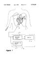
- FIG. 1 illustrates a functional block diagram for an ultrasound imaging machine.
- FIG. 2 illustrates a process flowchart in time for the method.
- FIG. 3 illustrates a variation of the flowchart shown in FIG. 2.
- FIG. 4 illustrates a variation of the flowchart shown in FIG. 2.
- FIG. 5 illustrates a variation of the flowchart shown in FIG. 2.
- FIG. 6 illustrates a variation of the flowchart shown in FIG. 2.
- FIG. 7 illustrates a variation of the flowchart shown in FIG. 6.
- FIG. 8 illustrates a variation of the flowchart shown in FIG. 2 and FIG. 3.
- FIG. 9 illustrates a variation of the flowchart shown in FIG. 8.
- FIG. 10 illustrates a variation of the flowchart shown in FIG. 2.
- FIG. 11 illustrates a variation of the flowchart shown in FIG. 2.
- FIG. 12 illustrates a variation of the flowchart shown in FIG. 10.
- FIG. 13 illustrates a variation of the flowchart shown in FIG. 12.
- Contrast agents resonate in the presence of an ultrasound field and the received signal can be detected by today's ultrasound scanners. It has been observed that higher transit powers increase the backscattering cross section of the microbubbles, particularly in harmonic mode, and they therefore scatter a larger portion of the impinging ultrasound signal. However, the duration of the contrast effect is shorter at high power levels, probably due to increased microbubble destruction and the subsequent lower concentrations. Different imaging sequences can be used to optimize this contrast effect.
- Synchronizing the imaging sequence to the patient's ECG allows a series of high-power frames to be strategically obtained. These frames may be fired at the same point of the heart cycle or at different points to allow for the piecing together of a complete heart cycle from multiple heart cycles using cine-loop technology. Minimizing high-power frames will allow for prolonged contrast effect thereby making endocardial border detection easier for ejection fraction and volume calculations as well as wall motion analysis. Simultaneously, the high-power frames make measurement of myocardial perfusion and critical timing parameters, such as wash-in and wash-out of contrast, possible. Gating off of a respiration signal will help minimize artifacts resulting from heart movement caused by patient breathing from heart cycle to heart cycle.
- the sequence may be accomplished with a mix of fundamental imaging and harmonic imaging. For example, it may be best to transmit the high-power frames in harmonic mode and the low-power frames in fundamental mode. These sequences are applicable across all ultrasound imaging modalities--2D imaging, Color Flow, Power Doppler Imaging, Doppler tissue colorization, etc.
- each ultrasound image frame is made up of 100 or more transmit lines which are steered at different angles. If fewer lines were transmitted it would be possible to use parallel processing on the receive path to fill an image. Combining this with the imaging sequences discussed will provide an even longer contrast effect.
- FIG. 1 illustrates a functional block diagram of an ultrasound imaging machine 110.
- An ultrasound probe 114 is connected to a receiving circuit 116 and a transmitting circuit 117.
- the receiving circuit 116 is further connected to a display 118.
- contrast agent is injected into a patient's bloodstream, such as the superior vena cava.
- the contrast agent improves the blood pool backscatter relative to the tissue.
- the blood is black and the tissue is white.
- ultrasound image energy is applied to the heart 112 via the ultrasound probe 114.
- the received images may be used characterize the cardiac blood flow of the patient.
- FIG. 2 illustrates a time process flowchart for the method.
- step 10 one high power frame per cycle is shot and the image is gathered.
- the high power frame is optionally triggered off of the ECG waveform or triggered off of the ECG and further gated by the respiration waveform.
- the high power frames may be processed separately and stored in a cine loop.
- FIG. 3 illustrates a variation of the process flowchart shown in FIG. 2.
- FIG. 4 illustrates another variation of the process flowchart shown in FIG. 2.
- ECG trigger i.e., R-wave
- FIG. 5 illustrates another variation of the process flowchart shown in FIG. 2.
- the frames can be delayed from an ECG trigger and gated by respiration. This is a special case of FIG. 3 without the low power frames.
- FIG. 6 illustrates another variation of the process flowchart shown in FIG. 2. This is a combination of the methods disclosed in FIGS. 2 and 4. It is possible that flow can be measured from this method.
- the second high power frame, shot every N (N> 1) cycles, but not necessarily the same cycle as the first high power frame, then measures the amount of contrast agent which has returned since the first high power frame. This sequence can give an indication of myocardial (or other organ) blood flow.
- FIG. 7 is a special case of FIG. 6 where only high power frames are shot.
- FIG. 8 illustrates a variation of FIGS. 2 and 3.
- FIG. 9 illustrates a variation of FIG. 8, with no low power frames being shot.
- FIG. 10 illustrates a variation of FIG. 2.
- the sequence is once again synchronized off of the ECG or ECG and gated by respiration.
- FIG. 11 illustrates a variation of FIG. 2.
- an unsynchronized frame rate control could be used with contrast agents, allowing dial-back from standard 30 Hz imaging.
- FIG. 12 is variation of FIG. 10.
- FIG. 13 is a variation of FIG. 12 with the low power frames not fired.
Abstract
Description
Claims (20)
Priority Applications (6)
| Application Number | Priority Date | Filing Date | Title |
|---|---|---|---|
| US08/695,267 US5735281A (en) | 1996-08-09 | 1996-08-09 | Method of enhancing and prolonging the effect of ultrasound contrast agents |
| DE19723053A DE19723053C2 (en) | 1996-08-09 | 1997-06-02 | Ultrasound imaging device |
| GB9714034A GB2316174B (en) | 1996-08-09 | 1997-07-02 | Improvements in ultrasound imaging |
| JP17961897A JP4465050B2 (en) | 1996-08-09 | 1997-07-04 | Ultrasonic imaging device |
| JP2007208929A JP4465373B2 (en) | 1996-08-09 | 2007-08-10 | Ultrasound imaging device |
| JP2009131850A JP4938051B2 (en) | 1996-08-09 | 2009-06-01 | Ultrasound imaging device |
Applications Claiming Priority (1)
| Application Number | Priority Date | Filing Date | Title |
|---|---|---|---|
| US08/695,267 US5735281A (en) | 1996-08-09 | 1996-08-09 | Method of enhancing and prolonging the effect of ultrasound contrast agents |
Publications (1)
| Publication Number | Publication Date |
|---|---|
| US5735281A true US5735281A (en) | 1998-04-07 |
Family
ID=24792321
Family Applications (1)
| Application Number | Title | Priority Date | Filing Date |
|---|---|---|---|
| US08/695,267 Expired - Lifetime US5735281A (en) | 1996-08-09 | 1996-08-09 | Method of enhancing and prolonging the effect of ultrasound contrast agents |
Country Status (4)
| Country | Link |
|---|---|
| US (1) | US5735281A (en) |
| JP (3) | JP4465050B2 (en) |
| DE (1) | DE19723053C2 (en) |
| GB (1) | GB2316174B (en) |
Cited By (56)
| Publication number | Priority date | Publication date | Assignee | Title |
|---|---|---|---|---|
| WO1998047533A1 (en) * | 1997-04-24 | 1998-10-29 | Nycomed Imaging A.S. | Ultrasound imaging of tissue perfusion by pulse energy disruption of contrast agent |
| US5833613A (en) * | 1996-09-27 | 1998-11-10 | Advanced Technology Laboratories, Inc. | Ultrasonic diagnostic imaging with contrast agents |
| WO1998053855A1 (en) * | 1997-05-30 | 1998-12-03 | Alliance Pharmaceutical Corp. | Methods and apparatus for monitoring and quantifying the movement of fluid |
| US5860931A (en) * | 1997-10-10 | 1999-01-19 | Acuson Corporation | Ultrasound method and system for measuring perfusion |
| WO1999008599A1 (en) * | 1997-08-21 | 1999-02-25 | Acuson Corporation | Ultrasonic method and system for imaging blood flow |
| US5882315A (en) * | 1997-12-23 | 1999-03-16 | Acuson Corporation | Ultrasonic imaging method and image for doppler tissue parameters |
| WO1999030619A1 (en) | 1997-12-17 | 1999-06-24 | Nycomed Imaging As | Ultrasonography of the prostate |
| US5935069A (en) * | 1997-10-10 | 1999-08-10 | Acuson Corporation | Ultrasound system and method for variable transmission of ultrasonic signals |
| US5957845A (en) * | 1997-04-11 | 1999-09-28 | Acuson Corporation | Gated ultrasound imaging apparatus and method |
| US6014473A (en) | 1996-02-29 | 2000-01-11 | Acuson Corporation | Multiple ultrasound image registration system, method and transducer |
| US6015384A (en) * | 1998-08-31 | 2000-01-18 | Acuson Corporation | Ultrasonic system and method for tissue viability imaging |
| US6045508A (en) * | 1997-02-27 | 2000-04-04 | Acuson Corporation | Ultrasonic probe, system and method for two-dimensional imaging or three-dimensional reconstruction |
| US6077225A (en) * | 1998-01-23 | 2000-06-20 | Hewlett-Packard Company | Ultrasound method for enhancing image presentation when contrast agents are used |
| US6080107A (en) * | 1999-01-26 | 2000-06-27 | Hewlett-Packard Company | Methods for the use of contrast agents in ultrasonic imaging |
| US6086540A (en) * | 1997-10-07 | 2000-07-11 | Molecular Biosystems, Inc. | Methods of ultrasound imaging using echogenically persistent contrast agents |
| US6149597A (en) * | 1997-11-26 | 2000-11-21 | Kabushiki Kaisha Toshiba | Diagnostic ultrasound imaging using contrast medium |
| US6171246B1 (en) | 1999-04-29 | 2001-01-09 | Michalakis Averkiou | Realtime ultrasonic imaging of perfusion using ultrasonic contrast agents |
| US6174287B1 (en) | 1999-06-11 | 2001-01-16 | Acuson Corporation | Medical diagnostic ultrasound system and method for continuous M-mode imaging and periodic imaging of contrast agents |
| US6210333B1 (en) | 1999-10-12 | 2001-04-03 | Acuson Corporation | Medical diagnostic ultrasound system and method for automated triggered intervals |
| US6224554B1 (en) * | 1999-03-31 | 2001-05-01 | Point Biomedical Corporation | Method to measure ambient fluid pressure |
| US6245019B1 (en) | 1998-05-11 | 2001-06-12 | Kabushiki Kaisha Toshiba | Ultrasonic diagnostic apparatus |
| US6258033B1 (en) | 1999-11-30 | 2001-07-10 | Agilent Technologies, Inc. | Ultrasound method employing echoes from a region of interest to enable quantization of backscatter signals |
| US6302846B1 (en) * | 1999-09-20 | 2001-10-16 | Acuson Corporation | Ultrasound method for assessing ejection fraction using ultrasound contrast agents |
| US6319203B1 (en) | 2000-07-28 | 2001-11-20 | Atl Ultrasound | Ultrasonic nonlinear imaging at fundamental frequencies |
| US6322512B1 (en) | 1997-01-22 | 2001-11-27 | Acuson Corporation | Ultrasound contrast imaging |
| EP1169966A1 (en) | 2000-07-06 | 2002-01-09 | Esaote S.p.A. | Method and apparatus for ultrasound imaging in the presence of contrast agents, particulary in the field of cardiology |
| US6340348B1 (en) | 1999-07-02 | 2002-01-22 | Acuson Corporation | Contrast agent imaging with destruction pulses in diagnostic medical ultrasound |
| US6436045B1 (en) | 2000-04-27 | 2002-08-20 | Koninklijke Phillips Electronics N.V. | User controlled destructive waveform routine for ultrasound systems |
| US6461303B2 (en) * | 2000-01-19 | 2002-10-08 | Bjorn Angelsen | Method of detecting ultrasound contrast agent in soft tissue, and quantitating blood perfusion through regions of tissue |
| US6464644B2 (en) | 2000-03-08 | 2002-10-15 | Ge Medical Systems Global Technology Company, Llc | Method of ultrasonic imaging and ultrasonic diagnostic apparatus |
| KR20020079560A (en) * | 2001-04-11 | 2002-10-19 | 지이 메디컬 시스템즈 글로발 테크놀러지 캄파니 엘엘씨 | Ultrasonic transmission/reception method, ultrasonic transmission/reception apparatus, ultrasonic imaging method and ultrasonic imaging apparatus |
| US6491633B1 (en) | 2000-03-10 | 2002-12-10 | Acuson Corporation | Medical diagnostic ultrasound system and method for contrast agent image beamformation |
| US6547738B2 (en) | 2001-05-03 | 2003-04-15 | Ge Medical Systems Global Technology Company, Llc | Methods and apparatus for using ultrasound with contrast agent |
| EP1314398A1 (en) * | 2001-11-22 | 2003-05-28 | Kabushiki Kaisha Toshiba | Ultrasonic diagnostic apparatus |
| US20040039286A1 (en) * | 2002-08-26 | 2004-02-26 | The Cleveland Clinic Foundation | System and method of aquiring blood-vessel data |
| US6726629B1 (en) | 1998-01-16 | 2004-04-27 | Acuson Corporation | Ultrasound contrast imaging |
| US20040133107A1 (en) * | 2002-11-01 | 2004-07-08 | Hiroshi Hashimoto | Ultrasonic diagnostic apparatus |
| US20040143241A1 (en) * | 2000-01-18 | 2004-07-22 | Joel Douglas | Subcutaneous injection set for use with a reservoir that has a septum |
| EP1514516A1 (en) * | 2003-09-11 | 2005-03-16 | Kabushiki Kaisha Toshiba | Ultrasonic diagnostic equipment and image processing apparatus |
| US20050245828A1 (en) * | 2004-04-20 | 2005-11-03 | Kabushiki Kaisha Toshiba | Ultrasound imaging apparatus and method of ultrasound imaging |
| US20050256404A1 (en) * | 2004-05-11 | 2005-11-17 | Kabushiki Kaisha Toshiba | Ultrasonic diagnostic apparatus and ultrasonic diagnostic method |
| US20050267371A1 (en) * | 2001-08-22 | 2005-12-01 | Yoichi Ogasawara | Ultrasonic diagnostic apparatus |
| USRE38971E1 (en) | 1995-01-31 | 2006-02-07 | Kabushiki Kaisha Toshiba | Ultrasound diagnostic apparatus and method |
| US20060155192A1 (en) * | 2002-11-29 | 2006-07-13 | Ragnar Bendiksen | Ultrasound triggering method |
| US20070078344A1 (en) * | 2003-10-23 | 2007-04-05 | Koninklijke Philips Electronics N.V. | Ultrasound imaging method and apparatus |
| CN100366225C (en) * | 2000-12-26 | 2008-02-06 | 株式会社东芝 | Ultrasonic diagnosis apparatus and control method of ultrasonic diagnosis apparatus |
| DE102006057211B3 (en) * | 2006-12-01 | 2008-03-27 | Kompetenzzentrum Medizintechnik Ruhr (Kmr) E.V. | Object e.g. biological tissue, infusion measuring method, involves determining re-enrichment conditions in time period, in which no tomogram series is received, by mathematical approximation technique |
| US20080255453A1 (en) * | 2007-04-13 | 2008-10-16 | Atsuko Matsunaga | Ultrasonic diagnostic apparatus |
| CN100518660C (en) * | 2004-03-24 | 2009-07-29 | 株式会社东芝 | Ultrasound diagnostic apparatus |
| CN101347343B (en) * | 2007-07-17 | 2010-12-22 | 株式会社东芝 | Apparatus of acquiring ultrasonic images |
| EP2644099A1 (en) * | 2012-03-28 | 2013-10-02 | Samsung Medison Co., Ltd. | Ultrasound system and method of obtaining ultrasound image |
| US20140357999A1 (en) * | 2013-05-29 | 2014-12-04 | B-K Medical Aps | Color flow ultrasound imaging |
| CN107822660A (en) * | 2016-07-26 | 2018-03-23 | 美国西门子医疗解决公司 | Generate method, ultrasonic system and the storage medium of ultrasonoscopy |
| US20180228554A1 (en) * | 2001-02-13 | 2018-08-16 | Mediguide Ltd. | System and method for delivering a stent to a selected position within a lumen |
| US11058397B2 (en) * | 2015-01-02 | 2021-07-13 | Esaote S.P.A. | Method for quantifying the elasticity of a material by ultrasounds |
| US11918418B1 (en) * | 2023-04-06 | 2024-03-05 | Orcasonics Innovation Ltd | Ultrasound imaging systems, devices and methods that simulate a virtual moving transducer/receiver to implement a synthetic aperture array |
Families Citing this family (5)
| Publication number | Priority date | Publication date | Assignee | Title |
|---|---|---|---|---|
| US6210335B1 (en) * | 1999-12-08 | 2001-04-03 | General Electric Company | Acoustic flash to increase penetration |
| JP4764125B2 (en) * | 2005-09-21 | 2011-08-31 | 株式会社東芝 | Ultrasonic diagnostic apparatus and control program for ultrasonic diagnostic apparatus |
| JP5162923B2 (en) * | 2007-02-27 | 2013-03-13 | 株式会社日立製作所 | Ultrasonic imaging device |
| WO2013116807A1 (en) | 2012-02-03 | 2013-08-08 | Los Alamos National Security, Llc | Systems and methods for synthetic aperture ultrasound tomography |
| WO2013116783A1 (en) | 2012-02-03 | 2013-08-08 | Los Alamos National Security, Llc | Windowed time-reversal music technique for super-resolution ultrasound imaging |
Citations (2)
| Publication number | Priority date | Publication date | Assignee | Title |
|---|---|---|---|---|
| US5410205A (en) * | 1993-02-11 | 1995-04-25 | Hewlett-Packard Company | Ultrasonic transducer having two or more resonance frequencies |
| US5577505A (en) * | 1996-02-06 | 1996-11-26 | Hewlett-Packard Company | Means for increasing sensitivity in non-linear ultrasound imaging systems |
Family Cites Families (14)
| Publication number | Priority date | Publication date | Assignee | Title |
|---|---|---|---|---|
| JPS58155846A (en) * | 1982-03-12 | 1983-09-16 | 富士通株式会社 | Ultrasonic contrast gas bubble generating method and apparatus |
| JPS6427540A (en) * | 1987-07-24 | 1989-01-30 | Hitachi Medical Corp | Ultrasonic diagnostic apparatus |
| JPS63255045A (en) * | 1987-09-28 | 1988-10-21 | 株式会社東芝 | Ultrasonic diagnostic apparatus |
| JPH01221144A (en) * | 1987-11-24 | 1989-09-04 | Hitachi Ltd | Ultrasonic measuring device |
| JPH01230346A (en) * | 1988-03-11 | 1989-09-13 | Hitachi Medical Corp | Diagnosis device by ultra sonic wave |
| US5016642A (en) * | 1990-04-11 | 1991-05-21 | Hewlett-Packard Company | Slow motion cardiac imaging |
| JP3096818B2 (en) * | 1990-10-12 | 2000-10-10 | 株式会社日立メディコ | Ultrasound diagnostic equipment |
| JPH0678926A (en) * | 1992-09-03 | 1994-03-22 | Toshiba Corp | Ultrasonic diagnostic device |
| US5456257A (en) * | 1994-11-23 | 1995-10-10 | Advanced Technology Laboratories, Inc. | Ultrasonic detection of contrast agents |
| JP3459304B2 (en) * | 1995-01-31 | 2003-10-20 | 株式会社東芝 | Ultrasound diagnostic equipment |
| JP3023290B2 (en) * | 1995-04-14 | 2000-03-21 | 株式会社東芝 | Ultrasound diagnostic equipment |
| JP3488541B2 (en) * | 1995-06-09 | 2004-01-19 | 株式会社東芝 | Ultrasound diagnostic equipment |
| JP3510025B2 (en) * | 1995-11-10 | 2004-03-22 | ジーイー横河メディカルシステム株式会社 | Ultrasound imaging device |
| JP3510032B2 (en) * | 1996-01-12 | 2004-03-22 | ジーイー横河メディカルシステム株式会社 | Ultrasound imaging device |
-
1996
- 1996-08-09 US US08/695,267 patent/US5735281A/en not_active Expired - Lifetime
-
1997
- 1997-06-02 DE DE19723053A patent/DE19723053C2/en not_active Expired - Lifetime
- 1997-07-02 GB GB9714034A patent/GB2316174B/en not_active Expired - Lifetime
- 1997-07-04 JP JP17961897A patent/JP4465050B2/en not_active Expired - Lifetime
-
2007
- 2007-08-10 JP JP2007208929A patent/JP4465373B2/en not_active Expired - Lifetime
-
2009
- 2009-06-01 JP JP2009131850A patent/JP4938051B2/en not_active Expired - Lifetime
Patent Citations (2)
| Publication number | Priority date | Publication date | Assignee | Title |
|---|---|---|---|---|
| US5410205A (en) * | 1993-02-11 | 1995-04-25 | Hewlett-Packard Company | Ultrasonic transducer having two or more resonance frequencies |
| US5577505A (en) * | 1996-02-06 | 1996-11-26 | Hewlett-Packard Company | Means for increasing sensitivity in non-linear ultrasound imaging systems |
Non-Patent Citations (2)
| Title |
|---|
| Circulation, vol. 92, No. 9, Nov. 1, 1995 Transient Mycardial Contrast After Initial Exposure to Diagnostic Ultrasound Pressures With Minute Doses of Intravenously Injected Microbubbles Thomas R. Porter, MD; Feng Xie, MD/pp. 2391 2395. * |
| Circulation, vol. 92, No. 9, Nov. 1, 1995 Transient Mycardial Contrast After Initial Exposure to Diagnostic Ultrasound Pressures With Minute Doses of Intravenously Injected Microbubbles Thomas R. Porter, MD; Feng Xie, MD/pp. 2391-2395. |
Cited By (92)
| Publication number | Priority date | Publication date | Assignee | Title |
|---|---|---|---|---|
| US7081092B2 (en) | 1994-09-28 | 2006-07-25 | Imcor Pharmaceutical Company | Methods and apparatus for monitoring and quantifying the movement of fluid |
| US20050049484A1 (en) * | 1994-09-28 | 2005-03-03 | Schutt Ernest G. | Methods and apparatus for monitoring and quantifying the movement of fluid |
| USRE38971E1 (en) | 1995-01-31 | 2006-02-07 | Kabushiki Kaisha Toshiba | Ultrasound diagnostic apparatus and method |
| US6575910B2 (en) | 1995-10-10 | 2003-06-10 | Koninklijke Philips Electronics N.V. | Ultrasonic image persistence using contrast agents |
| US6540684B2 (en) | 1995-10-10 | 2003-04-01 | Koninklijke Philips Electronics N.V. | Ultrasonic perfusion measurement using contrast agents |
| US6360027B1 (en) * | 1996-02-29 | 2002-03-19 | Acuson Corporation | Multiple ultrasound image registration system, method and transducer |
| US6132376A (en) * | 1996-02-29 | 2000-10-17 | Acuson Corporation | Multiple ultrasonic image registration system, method and transducer |
| US6201900B1 (en) | 1996-02-29 | 2001-03-13 | Acuson Corporation | Multiple ultrasound image registration system, method and transducer |
| US6222948B1 (en) | 1996-02-29 | 2001-04-24 | Acuson Corporation | Multiple ultrasound image registration system, method and transducer |
| US6014473A (en) | 1996-02-29 | 2000-01-11 | Acuson Corporation | Multiple ultrasound image registration system, method and transducer |
| US6102865A (en) * | 1996-02-29 | 2000-08-15 | Acuson Corporation | Multiple ultrasound image registration system, method and transducer |
| US5833613A (en) * | 1996-09-27 | 1998-11-10 | Advanced Technology Laboratories, Inc. | Ultrasonic diagnostic imaging with contrast agents |
| US6322512B1 (en) | 1997-01-22 | 2001-11-27 | Acuson Corporation | Ultrasound contrast imaging |
| US6171248B1 (en) | 1997-02-27 | 2001-01-09 | Acuson Corporation | Ultrasonic probe, system and method for two-dimensional imaging or three-dimensional reconstruction |
| US6045508A (en) * | 1997-02-27 | 2000-04-04 | Acuson Corporation | Ultrasonic probe, system and method for two-dimensional imaging or three-dimensional reconstruction |
| US5957845A (en) * | 1997-04-11 | 1999-09-28 | Acuson Corporation | Gated ultrasound imaging apparatus and method |
| US6306095B1 (en) | 1997-04-11 | 2001-10-23 | Acuson Corporation | Gated ultrasound imaging apparatus and method |
| US6626831B2 (en) | 1997-04-11 | 2003-09-30 | Acuson Corporation | Gated ultrasound imaging apparatus and method |
| US6315730B1 (en) | 1997-04-24 | 2001-11-13 | Nyomed Imaging As | Ultrasound imaging of tissue perfusion by pulse energy disruption of contrast agent |
| WO1998047533A1 (en) * | 1997-04-24 | 1998-10-29 | Nycomed Imaging A.S. | Ultrasound imaging of tissue perfusion by pulse energy disruption of contrast agent |
| US6802813B2 (en) | 1997-05-30 | 2004-10-12 | Ernest G. Schutt | Methods and apparatus for monitoring and quantifying the movement of fluid |
| WO1998053855A1 (en) * | 1997-05-30 | 1998-12-03 | Alliance Pharmaceutical Corp. | Methods and apparatus for monitoring and quantifying the movement of fluid |
| US5947904A (en) * | 1997-08-21 | 1999-09-07 | Acuson Corporation | Ultrasonic method and system for imaging blood flow including disruption or activation of a contrast agent |
| US5944666A (en) * | 1997-08-21 | 1999-08-31 | Acuson Corporation | Ultrasonic method for imaging blood flow including disruption or activation of contrast agent |
| WO1999008599A1 (en) * | 1997-08-21 | 1999-02-25 | Acuson Corporation | Ultrasonic method and system for imaging blood flow |
| US6086540A (en) * | 1997-10-07 | 2000-07-11 | Molecular Biosystems, Inc. | Methods of ultrasound imaging using echogenically persistent contrast agents |
| US5935069A (en) * | 1997-10-10 | 1999-08-10 | Acuson Corporation | Ultrasound system and method for variable transmission of ultrasonic signals |
| US5860931A (en) * | 1997-10-10 | 1999-01-19 | Acuson Corporation | Ultrasound method and system for measuring perfusion |
| US6149597A (en) * | 1997-11-26 | 2000-11-21 | Kabushiki Kaisha Toshiba | Diagnostic ultrasound imaging using contrast medium |
| US6689065B2 (en) * | 1997-12-17 | 2004-02-10 | Amersham Health As | Ultrasonography |
| WO1999030619A1 (en) | 1997-12-17 | 1999-06-24 | Nycomed Imaging As | Ultrasonography of the prostate |
| US5882315A (en) * | 1997-12-23 | 1999-03-16 | Acuson Corporation | Ultrasonic imaging method and image for doppler tissue parameters |
| US6110117A (en) * | 1997-12-23 | 2000-08-29 | Acuson Corporation | Ultrasonic imaging method and image for doppler tissue parameters |
| US6726629B1 (en) | 1998-01-16 | 2004-04-27 | Acuson Corporation | Ultrasound contrast imaging |
| US6077225A (en) * | 1998-01-23 | 2000-06-20 | Hewlett-Packard Company | Ultrasound method for enhancing image presentation when contrast agents are used |
| US6245019B1 (en) | 1998-05-11 | 2001-06-12 | Kabushiki Kaisha Toshiba | Ultrasonic diagnostic apparatus |
| US6015384A (en) * | 1998-08-31 | 2000-01-18 | Acuson Corporation | Ultrasonic system and method for tissue viability imaging |
| US6080107A (en) * | 1999-01-26 | 2000-06-27 | Hewlett-Packard Company | Methods for the use of contrast agents in ultrasonic imaging |
| US6224554B1 (en) * | 1999-03-31 | 2001-05-01 | Point Biomedical Corporation | Method to measure ambient fluid pressure |
| US6171246B1 (en) | 1999-04-29 | 2001-01-09 | Michalakis Averkiou | Realtime ultrasonic imaging of perfusion using ultrasonic contrast agents |
| US6174287B1 (en) | 1999-06-11 | 2001-01-16 | Acuson Corporation | Medical diagnostic ultrasound system and method for continuous M-mode imaging and periodic imaging of contrast agents |
| US6340348B1 (en) | 1999-07-02 | 2002-01-22 | Acuson Corporation | Contrast agent imaging with destruction pulses in diagnostic medical ultrasound |
| US6302846B1 (en) * | 1999-09-20 | 2001-10-16 | Acuson Corporation | Ultrasound method for assessing ejection fraction using ultrasound contrast agents |
| US6210333B1 (en) | 1999-10-12 | 2001-04-03 | Acuson Corporation | Medical diagnostic ultrasound system and method for automated triggered intervals |
| US6258033B1 (en) | 1999-11-30 | 2001-07-10 | Agilent Technologies, Inc. | Ultrasound method employing echoes from a region of interest to enable quantization of backscatter signals |
| US20040143241A1 (en) * | 2000-01-18 | 2004-07-22 | Joel Douglas | Subcutaneous injection set for use with a reservoir that has a septum |
| US6461303B2 (en) * | 2000-01-19 | 2002-10-08 | Bjorn Angelsen | Method of detecting ultrasound contrast agent in soft tissue, and quantitating blood perfusion through regions of tissue |
| US6464644B2 (en) | 2000-03-08 | 2002-10-15 | Ge Medical Systems Global Technology Company, Llc | Method of ultrasonic imaging and ultrasonic diagnostic apparatus |
| US6491633B1 (en) | 2000-03-10 | 2002-12-10 | Acuson Corporation | Medical diagnostic ultrasound system and method for contrast agent image beamformation |
| US6436045B1 (en) | 2000-04-27 | 2002-08-20 | Koninklijke Phillips Electronics N.V. | User controlled destructive waveform routine for ultrasound systems |
| EP1169966A1 (en) | 2000-07-06 | 2002-01-09 | Esaote S.p.A. | Method and apparatus for ultrasound imaging in the presence of contrast agents, particulary in the field of cardiology |
| US6561982B2 (en) | 2000-07-06 | 2003-05-13 | Esaote, S.P.A. | Method and apparatus for ultrasound imaging in the presence of contrast agents, Particularly in the field of cardiology |
| US6319203B1 (en) | 2000-07-28 | 2001-11-20 | Atl Ultrasound | Ultrasonic nonlinear imaging at fundamental frequencies |
| US6544182B2 (en) | 2000-07-28 | 2003-04-08 | Koninklijke Philips Electronics N.V. | Ultrasonic nonlinear imaging at fundamental frequencies |
| CN100366225C (en) * | 2000-12-26 | 2008-02-06 | 株式会社东芝 | Ultrasonic diagnosis apparatus and control method of ultrasonic diagnosis apparatus |
| US20180228554A1 (en) * | 2001-02-13 | 2018-08-16 | Mediguide Ltd. | System and method for delivering a stent to a selected position within a lumen |
| KR20020079560A (en) * | 2001-04-11 | 2002-10-19 | 지이 메디컬 시스템즈 글로발 테크놀러지 캄파니 엘엘씨 | Ultrasonic transmission/reception method, ultrasonic transmission/reception apparatus, ultrasonic imaging method and ultrasonic imaging apparatus |
| US6547738B2 (en) | 2001-05-03 | 2003-04-15 | Ge Medical Systems Global Technology Company, Llc | Methods and apparatus for using ultrasound with contrast agent |
| US20050267371A1 (en) * | 2001-08-22 | 2005-12-01 | Yoichi Ogasawara | Ultrasonic diagnostic apparatus |
| EP1314398A1 (en) * | 2001-11-22 | 2003-05-28 | Kabushiki Kaisha Toshiba | Ultrasonic diagnostic apparatus |
| US6641538B2 (en) | 2001-11-22 | 2003-11-04 | Kabushiki Kaisha Toshiba | Ultrasonic diagnostic apparatus and method of controlling a ultrasonic diagnostic apparatus |
| US20040039286A1 (en) * | 2002-08-26 | 2004-02-26 | The Cleveland Clinic Foundation | System and method of aquiring blood-vessel data |
| US20110208017A1 (en) * | 2002-08-26 | 2011-08-25 | Kuban Barry D | System and method of aquiring blood-vessel data |
| US8622910B2 (en) | 2002-08-26 | 2014-01-07 | The Cleveland Clinic Foundation | System and method of aquiring blood-vessel data |
| US7927275B2 (en) | 2002-08-26 | 2011-04-19 | The Cleveland Clinic Foundation | System and method of aquiring blood-vessel data |
| US6786869B2 (en) | 2002-11-01 | 2004-09-07 | Ge Medical Systems Global Technology Company, Llc | Ultrasonic diagnostic apparatus |
| US20040133107A1 (en) * | 2002-11-01 | 2004-07-08 | Hiroshi Hashimoto | Ultrasonic diagnostic apparatus |
| US20060155192A1 (en) * | 2002-11-29 | 2006-07-13 | Ragnar Bendiksen | Ultrasound triggering method |
| US7931595B2 (en) * | 2002-11-29 | 2011-04-26 | Ge Healthcare As | Ultrasound triggering method to reduce cardiac arrhythmia |
| EP1514516A1 (en) * | 2003-09-11 | 2005-03-16 | Kabushiki Kaisha Toshiba | Ultrasonic diagnostic equipment and image processing apparatus |
| US7588538B2 (en) * | 2003-09-11 | 2009-09-15 | Kabushiki Kaisha Toshiba | Ultrasonic diagnostic equipment and image processing apparatus |
| CN100457045C (en) * | 2003-09-11 | 2009-02-04 | 株式会社东芝 | Ultrasonic diagnostic equipment and image processing apparatus |
| US20050059893A1 (en) * | 2003-09-11 | 2005-03-17 | Yoichi Ogasawara | Ultrasonic dignostic equipment and image processing apparatus |
| US20070078344A1 (en) * | 2003-10-23 | 2007-04-05 | Koninklijke Philips Electronics N.V. | Ultrasound imaging method and apparatus |
| US7862511B2 (en) | 2003-10-23 | 2011-01-04 | Koninkliljke Philips Electronics N.V. | Ultrasound imaging method and apparatus |
| CN100518660C (en) * | 2004-03-24 | 2009-07-29 | 株式会社东芝 | Ultrasound diagnostic apparatus |
| US20050245828A1 (en) * | 2004-04-20 | 2005-11-03 | Kabushiki Kaisha Toshiba | Ultrasound imaging apparatus and method of ultrasound imaging |
| US8926516B2 (en) * | 2004-04-20 | 2015-01-06 | Kabushiki Kaisha Toshiba | Ultrasound imaging apparatus and method of ultrasound imaging |
| US8454517B2 (en) * | 2004-05-11 | 2013-06-04 | Kabushiki Kaisha Toshiba | Ultrasonic diagnostic apparatus and ultrasonic diagnostic method |
| US20050256404A1 (en) * | 2004-05-11 | 2005-11-17 | Kabushiki Kaisha Toshiba | Ultrasonic diagnostic apparatus and ultrasonic diagnostic method |
| WO2008064645A3 (en) * | 2006-12-01 | 2008-08-21 | Kompetenzzentrum Medizintechni | Method and device for measuring perfusion using a contrast medium |
| WO2008064645A2 (en) * | 2006-12-01 | 2008-06-05 | Kompetenzzentrum Medizintechnik Ruhr (Kmr) E.V. Ruhr Universität Bochum | Method and device for measuring perfusion using a contrast medium |
| DE102006057211B3 (en) * | 2006-12-01 | 2008-03-27 | Kompetenzzentrum Medizintechnik Ruhr (Kmr) E.V. | Object e.g. biological tissue, infusion measuring method, involves determining re-enrichment conditions in time period, in which no tomogram series is received, by mathematical approximation technique |
| US20080255453A1 (en) * | 2007-04-13 | 2008-10-16 | Atsuko Matsunaga | Ultrasonic diagnostic apparatus |
| CN101347343B (en) * | 2007-07-17 | 2010-12-22 | 株式会社东芝 | Apparatus of acquiring ultrasonic images |
| EP2644099A1 (en) * | 2012-03-28 | 2013-10-02 | Samsung Medison Co., Ltd. | Ultrasound system and method of obtaining ultrasound image |
| US9192360B2 (en) | 2012-03-28 | 2015-11-24 | Samsung Medison Co., Ltd. | Ultrasound system and method of obtaining ultrasound image |
| US20140357999A1 (en) * | 2013-05-29 | 2014-12-04 | B-K Medical Aps | Color flow ultrasound imaging |
| US9943289B2 (en) * | 2013-05-29 | 2018-04-17 | B-K Medical Aps | Color flow ultrasound imaging |
| US11058397B2 (en) * | 2015-01-02 | 2021-07-13 | Esaote S.P.A. | Method for quantifying the elasticity of a material by ultrasounds |
| CN107822660A (en) * | 2016-07-26 | 2018-03-23 | 美国西门子医疗解决公司 | Generate method, ultrasonic system and the storage medium of ultrasonoscopy |
| US11918418B1 (en) * | 2023-04-06 | 2024-03-05 | Orcasonics Innovation Ltd | Ultrasound imaging systems, devices and methods that simulate a virtual moving transducer/receiver to implement a synthetic aperture array |
Also Published As
| Publication number | Publication date |
|---|---|
| JP4465373B2 (en) | 2010-05-19 |
| DE19723053A1 (en) | 1998-02-12 |
| JP2009189867A (en) | 2009-08-27 |
| DE19723053C2 (en) | 2001-07-19 |
| GB2316174A (en) | 1998-02-18 |
| JPH1075951A (en) | 1998-03-24 |
| JP2007330804A (en) | 2007-12-27 |
| JP4465050B2 (en) | 2010-05-19 |
| GB9714034D0 (en) | 1997-09-10 |
| JP4938051B2 (en) | 2012-05-23 |
| GB2316174B (en) | 2000-06-28 |
Similar Documents
| Publication | Publication Date | Title |
|---|---|---|
| US5735281A (en) | Method of enhancing and prolonging the effect of ultrasound contrast agents | |
| US6110120A (en) | Gated ultrasound imaging apparatus and method | |
| US5846202A (en) | Ultrasound method and system for imaging | |
| US7862511B2 (en) | Ultrasound imaging method and apparatus | |
| US6340348B1 (en) | Contrast agent imaging with destruction pulses in diagnostic medical ultrasound | |
| US6858011B2 (en) | Method and apparatus to control microbubble destruction during contrast-enhanced ultrasound imaging, and uses therefor | |
| US5709210A (en) | Ultrasound system for imaging | |
| CN100518657C (en) | Contrast agent imaging with synchronized persistence | |
| Becher et al. | Harmonic power Doppler contrast echocardiography: preliminary clinical results | |
| JP2005512650A (en) | Ultrasound imaging system and method for displaying tissue perfusion and other parameters that change over time | |
| US20040087858A1 (en) | Method and apparatus for improving contrast-to-tissue ratio in ultrasound contrast imaging with subharmonic imaging | |
| US8926516B2 (en) | Ultrasound imaging apparatus and method of ultrasound imaging | |
| AU2003302593B2 (en) | Ultrasound triggering method | |
| US6730036B2 (en) | Ultrasonic imaging to detect coronary artery stenosis at rest | |
| US20090124908A1 (en) | Method And Apparatus For Detecting Ultrasound Contrast Agents In Arterioles | |
| US6302846B1 (en) | Ultrasound method for assessing ejection fraction using ultrasound contrast agents | |
| Gardner et al. | Synchronization of contrast agent destruction and imaging for perfusion assessment |
Legal Events
| Date | Code | Title | Description |
|---|---|---|---|
| AS | Assignment |
Owner name: HEWLETT-PACKARD COMPANY, CALIFORNIA Free format text: ASSIGNMENT OF ASSIGNORS INTEREST;ASSIGNORS:RAFTER, PATRICK G.;GATZKE, RONALD D.;D'SA ALWYN P.;REEL/FRAME:008138/0785 Effective date: 19960809 |
|
| STCF | Information on status: patent grant |
Free format text: PATENTED CASE |
|
| AS | Assignment |
Owner name: HEWLETT-PACKARD COMPANY, A DELAWARE CORPORATION, C Free format text: MERGER;ASSIGNOR:HEWLETT-PACKARD COMPANY, A CALIFORNIA CORPORATION;REEL/FRAME:010841/0649 Effective date: 19980520 |
|
| AS | Assignment |
Owner name: AGILENT TECHNOLOGIES INC, CALIFORNIA Free format text: ASSIGNMENT OF ASSIGNORS INTEREST;ASSIGNOR:HEWLETT-PACKARD COMPANY;REEL/FRAME:010977/0540 Effective date: 19991101 |
|
| FPAY | Fee payment |
Year of fee payment: 4 |
|
| AS | Assignment |
Owner name: KONINKLIJKE PHILIPS ELECTRONICS N.V., NETHERLANDS Free format text: ASSIGNMENT OF ASSIGNORS INTEREST;ASSIGNOR:AGILENT TECHNOLOGIES, INC.;REEL/FRAME:014662/0179 Effective date: 20010801 |
|
| FPAY | Fee payment |
Year of fee payment: 8 |
|
| AS | Assignment |
Owner name: KONINKLIJKE PHILIPS ELECTRONICS N V, NETHERLANDS Free format text: ASSIGNMENT OF ASSIGNORS INTEREST;ASSIGNOR:AGILENT TECHNOLOGIES, INC.;REEL/FRAME:022835/0572 Effective date: 20090610 |
|
| FPAY | Fee payment |
Year of fee payment: 12 |